Nutcracker syndrome
Images

Case Summary
A 21-year-old woman with no significant prior medical history was evaluated in the emergency room after being involved in a motor vehicle accident. Contrast-enhanced computed tomography (CT) of the chest, abdomen, and pelvis was performed to assess for trauma-related injuries. There were no trauma-related injuries. However, incidentally an abnormal-appearing left renal vein was observed. Urinalysis revealed proteinuria and hematuria. The patient denied any previous abdominal or pelvic symptoms, including pain.
Imaging Findings
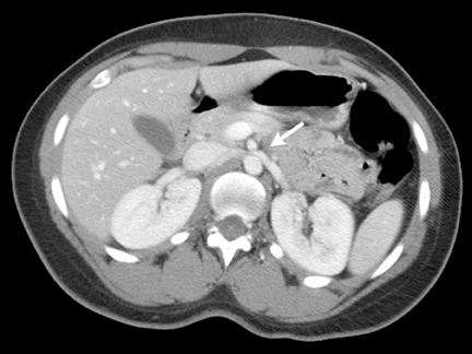
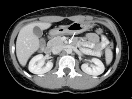
Axial contrast-enhanced CT of the abdomen demonstrated compression of the left renal vein between the superior mesenteric artery (SMA) and the aorta as the left renal vein crosses midline (Figure 1). An axial image of the same patient a few centimeters caudad demonstrated distended collateral vessels secondary to left renal vein compression adjacent to the renal hilum (Figure 2). Axial contrast-enhanced CT demonstrated left renal vein in a normal patient (without nutcracker syndrome) as it crosses between the SMA and the abdominal aorta (Figure 3).
Diagnosis
Nutcracker syndrome (renal vein entrapment)
Discussion
Nutcracker syndrome refers to the compression of the left renal vein between the aorta and the superior mesenteric artery, which results in elevated left renal vein pressure and possible collateral vein development. Clinically, Nutcracker syndrome is characterized by intermittent hematuria with or without left flank or abdominal pain. The syndrome occurs in relatively thin patients and adolescents who often have an otherwise healthy medical history.1 The true prevalence of Nutcracker syndrome remains unknown.
Nutcracker syndrome can have several clinical manifestations. The most common presentation is hematuria. Hematuria from the left ureteral orifice on cystoscopic examination in the absence of any detectable abnormality of the urinary tract should raise suspicion for Nutcracker syndrome.2 Nutcracker syndrome can also cause mild to moderate proteinuria. Other presentations that are rare include gonadal vein syndrome and varicocele.1,3,4 Prominent collateral vessels may develop, and the gonadal, ascending lumbar, adrenal, periureteral, and capsular veins are major potential collateral veins that can develop from left renal vein compression or obstruction.1
The mechanism for producing hematuria is thought to be due to increased left renal vein pressure, resulting in small venous ruptures into the collecting system or between dilated venous sinuses and adjacent renal calyces.2 Nutcracker syndrome should be part of the differential consideration in the evaluation for hematuria when other etiologies have been excluded.5
The aortomesenteric space is normally quite wide but in patients with nutcracker syndrome, it usually measures between 4 and 5 mm in width.2 The normally wide aortomesenteric angle is maintained by retroperitoneal fat and the third portion of the duodenum. A narrow aortomesenteric angle causes compression or entrapment of the left renal vein.2 A hypothesis for the narrowing of the aortomesenteric angle is a thin body habitus with decreased retroperitoneal and mesenteric fat. Other etiologic hypotheses of nutcracker syndrome include posterior renal ptosis with resultant stretching of the left renal vein over the aorta, and abnormal branching of the superior mesenteric artery from the aorta.6
Controversy exists about obtaining a reliable diagnosis of nutcracker syndrome. Although it can be invasive and uncomfortable for the patient, renal venography combined with measurement of the pressure gradient between the left renal vein and the IVC is the gold standard for demonstrating renal vein hypertension. However, no clear consensus exists on the cutoff of pressure gradient with which nutcracker syndrome can be clearly diagnosed.1
Doppler ultrasound measurements of the anterior-posterior (A-P) diameter and peak velocities of the left renal vein may be helpful in diagnosing nutcracker syndrome.1 Another study showed that correlation of renocaval pressure gradients with flow patterns from color Doppler sonography in collateral vessels further aids assessment of nutcracker syndrome.2
CT and CT angiography are other noninvasive modalities that can demonstrate compression of the left renal vein in the aortomesenteric angle and collateral veins. However, unlike Doppler sonography, flow characteristic cannot be made in collateral vessels. Magnetic resonance imaging (MRI) and MR angiography may also demonstrate the compression of the left renal vein between the superior mesenteric artery and the aorta.
Frequently, intravenous pyelogram and retrograde pyelographic studies are normal. The most common abnormal finding is ureteric or pelvic notching due to extrinsic compression from collateral vessels.
A confounding factor is that distended left renal vein can be a normal variant without collateral veins and with normal pressure gradient. Distinguishing between distended left renal veins that are a normal variant and those that indicate early nutcracker syndrome is difficult in patients with borderline left renal vein hypertension.2
Controversy also exists regarding treatment of nutcracker syndrome. Conservative management with routine urinalysis is proposed for mild hematuria, since the development of collateral veins may resolve the hypertension in the left renal vein and alleviate symptoms.6 Indications for surgery include severe persistent or recurrent hematuria causing anemia, and blood clots causing abdominal or flank pain. Surgical options include nephrectomy, variceal ligation, nephropexy, and renocaval reimplantation.7 More recently, endovascular treatment options have been applied.1,6,7
Conclusion
Nutcracker syndrome refers to the entrapment of the left renal vein in the mesoaortic angle with elevation of left renal vein pressure. Patients may present with hematuria, proteinuria, collateral vessel formation and pain. The gold standard for diagnosis of renal vein hypertension is renal venography combined with measurement of renocaval pressure gradient, although there is no consensus on criteria for a clear diagnosis. Color Doppler ultrasound, CT, and MRI can be used to help make the diagnosis.
References
- Kim S, et al. Nutcracker syndrome: Diagnosis with Doppler US. Radiology. 1996;989:93-97.
- Takebayashi S, et al. Diagnosis of the nutcracker syndrome with color Doppler sonography: Correlation with flow patterns on retrograde left renal venography. AJR Am J Roentgenol. 1999;72:39-43.
- Dogra V and Bhatt S. Acute painful scrotum. Radiol Clin N Am. 2004;42:349-363.
- Umeoka S, et al. Vascular dilatation in the pelvis: Identification with CT and MR imaging. Radiographics. 2004; 24: 193-208.
- Pan C. Evaluation of gross hematuria. Pediatr Clin N Am. 2006; 53:401-412.
- Hokama A and Oshiro Y. A thin 43-year-old woman with gross hematuria. Can Med Assoc J. 2005; 173: 251.
- Rudloff U, et al. Mesoaortic compression of the left renal vein (nutcracker syndrome): Case reports and review of the literature. Ann Vasc Surg. 2006;20:120-129.
Related Articles
Citation
VS TPBSD. Nutcracker syndrome. Appl Radiol. 2012;(11):36A-36C.
November 7, 2012