PACS analysis, processing, and reporting tools compete at ISCT Workstation Face-Off
June 19, 2013 – Volumetric imaging, automated segmentation, lesion measurement, and structured reporting were some of the key PACS functionalities physician-operators demonstrated at the 11th Annual Workstation Face-Off. The Face-off was held on June 18, 2013 as part of the 15th Annual International Symposium on Multidetector-Row CT hosted by the International Society of Computed Tomography (ISCT) from June 17-20 in Washington, DC.
For the past 10 years, the MDCT Workstation Faceoff has aimed to define the limits of workstation performance. Each year radiologists use advanced workstations to process and interpret several complex CT studies. Physician-operators navigate the same diverse clinical datasets to identify key clinical findings. Radiology workstations from the major manufacturers, including Carestream Health, GE Healthcare, Philips, Siemens, TeraRecon, and Vital Images, were tested before an audience of sophisticated physician judges under severe time pressure to demonstrate how the workstations and associated image reconstruction software can handle anything our moderator will throw at them.
This year—for the first time in its 10-year history—the competition required workstations that can analyze, automatically process, and report serial studies from multiple modalities. The exams included longitudinal examination of lung disease, liver tumor tracking, cardiac functional analysis, and neurovascularassessment.
“Efficient reading requires PACS workstations that can seamlessly handle multiple modalities, and offer advanced and intuitive tools to speed-up the assessment of complex cases. This enhances overall productivity and can reduce the need to resort to dedicated processing workstations that are not fully integrated in the reading workflow,” said Dr. Menashe Benjamin, Vice President, Healthcare Information Solutions at Carestream.
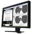
Carestream Health showcased the CARESTREAM Vue PACS workstation’s ability to support the proficient reading, processing and reporting of imaging studies from multiple modalities.
Radiologist Michalle Soudack, M.D., Head of Pediatric Radiology at the Safra Children’s Hospital in Israel, presented the cases for Carestream at the event. In presenting the exams, Dr. Soudack utilized Carestream’s new bookmarking capabilities. “Caretream’s advanced bookmarking allows me to quickly record findings of any type. Once stored in the PACS, the bookmarks can be utilized to easily navigate between findings over multiple studies and easily create comparisons. This can help speed up the (currently time consuming) reporting of longitudinal studies and possibly reduce the risk of human errors.”
According to Dr. Benjamin, “The integrated processing and reporting capabilities of the Vue PACS workstation were well depicted in this year’s Face-Off cases. For example, the Liver Tumor Tracking case involved following a target liver lesion over 2 time points. The recent study was a PET/CT while the prior exam was a CT.”
Carestream’s real-time registration capabilities synchronized the two CT series with the PET images. The company’s PET/CT and lesion management applications semi-automatically segmented the lesion in both time points and computed the RECIST measurements, 3D volume and maximal Standard Uptake Values (SUV) of the lesion. To complete the workflow, the application generated a quantitative report with tables and graphs of over-time information to facilitate assessment of the disease progression.
Another example was the neurovascular assessment case, involving studies from CTA, MRA and Rotational Angiogram modalities. The goal was to identify and assess the size of stenosis and aneurysm in the patient’s vertebrobasilar artery. “Carestream’s automatic registration feature was used to match the cross-modality studies, and to identify and display the findings. Our vessel analysis application was used to segment the vessel and to measure both the stenosis and the aneurysm,” Dr. Benjamin explained.
For more information: www.carestream.com, www.isct.org and www.appliedradiology.com
Related Articles
Citation
PACS analysis, processing, and reporting tools compete at ISCT Workstation Face-Off. Appl Radiol.
June 19, 2013
