Study Validates 3D Auto Segmentation MRI Tool for Vestibular Schwannoma
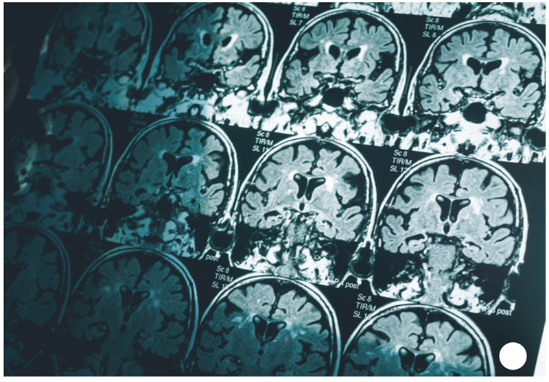 Researchers report that a deep convolutional neural networks (CNN) model delineated vestibular schwannoma using MRI with excellent accuracy, similar to human performance in most cases. The study, published in Radiology: Artificial Intelligence, found that the CNN model detected and delineated vestibular schwannomas accurately on contrast-enhanced T1- and T2-weighted MRI scans. It also distinguished the clinically relevant difference between intrameatal and extrameatal tumor parts.
The CNN model used in the study was a 3D U-Net with five encoder and decoder layers and trained as a multiclass segmentation task to automatically segment both the intra- and extrameatal component of the tumor. It was trained using fivefold cross-validation for two models (T1 and T2).
Vestibular schwannomas are rare, benign intracranial tumors arising from the neurilemma of the vestibular nerve. Initial symptoms usually comprise hearing loss, tinnitus, and balance disturbance.
In the study, MRI data from 214 patients in 37 different centers were retrospectively analyzed between 2020 and 2021. Of these, 134 positive for vestibular schwannoma [mean age 6 SD, 54 years 6 12; 64 men] and 80 negative for vestibular schwannoma were randomly assigned to a training and validation set and to an independent test set. To compare the computer and the human delineations, the authors used quantitative analysis, including Dice index, Hausdorff distance, surface-to-surface distance (S2S), and relative volume error.
The T1-weighted model showed state-of-the-art performance, with a mean S2S distance of less than 0.6 mm for the whole tumor and the intrameatal and extrameatal tumor parts. The whole tumor Dice index and Hausdorff distance were 0.92 and 2.1 mm in the independent test set, respectively. T2-weighted images had a mean S2S distance less than 0.6 mm for the whole tumor and the intrameatal and extrameatal tumor parts. The whole tumor Dice index and Hausdorff distance were 0.87 and 1.5 mm in the independent test set. The observer study indicated that the tool was similar to human delineations in 85%–92% of cases. The CNN detected tumors with 100% sensitivity and 99.1% specificity for the validation set and 100% sensitivity and 100% specificity for the test set.
The authors wrote, “the study shows the feasibility of automatically detecting and evaluating vestibular schwannoma with or without contrast material administration in large datasets acquired from multiple medical centers and MRI vendors.”