Study: Use of L1 Vertebral Level Expands Reach of CT Screening for Sarcopenia
 The use of the L1 vertebral level, typically included on both chest and abdominal CT examinations, instead of the L3 level expands the reach of opportunistic CT screening for sarcopenia, according to a study in the American Journal of Roentgenology.
“Automated CT-based measurements of muscle attenuation for myosteatosis at the L1 level compare favorably with previously-established L3 level measurements and clinical risk scores for predicting hip fractures and death,” wrote lead investigator Perry J. Pickhardt, MD, professor of radiology and chief, gastrointestinal imaging at the University of Wisconsin School of Medicine & Public Health in Madison. “Assessment for myopenia was less predictive of outcomes at both levels.”
Pickhardt and colleagues’ retrospective study included 9,223 patients (4,071 men, 5,152 women; mean age, 57 years) who underwent unenhanced low-dose abdominal CT. Muscle assessment for myosteatosis (mean attenuation) and myopenia (cross-sectional area) at the L1 and L3 levels was done via a previously validated fully-automated deep learning tool. Performance for predicting hip fractures and death was compared between L1 and L3 measures, then evaluated using FRAX score and Framingham risk score, respectively.
Among the 9,223 asymptomatic adults who underwent abdominal CT, muscle attenuation measurements at the L1 and L3 levels showed similar utility in predicting subsequent hip fracture and death. Meanwhile, muscle attenuation measurements at both levels showed comparable performance as established clinical risk scores.
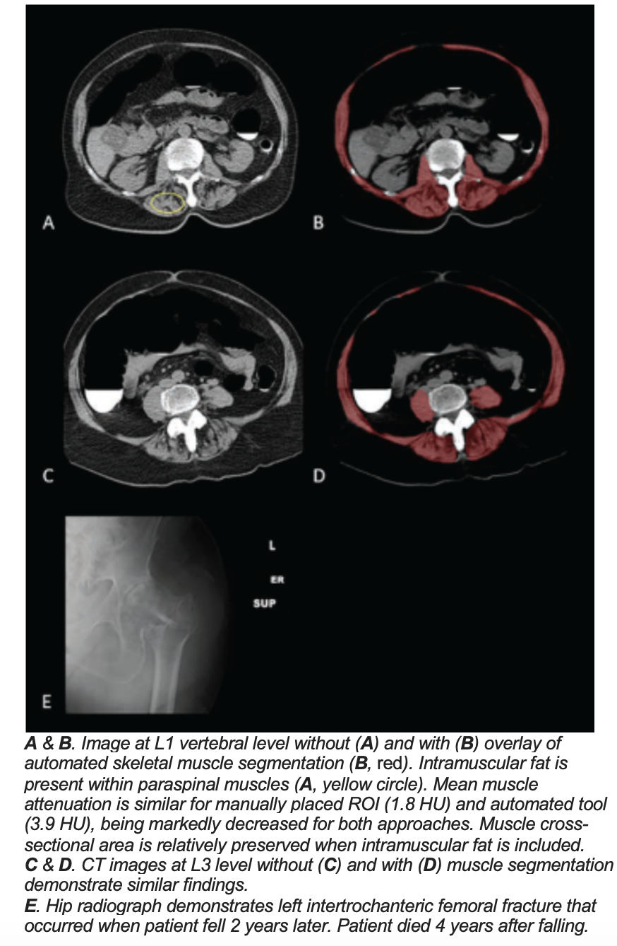
The assessment of muscle attenuation (myosteatosis) performed better than the assessment of muscle area (myopenia) “likely due in part to the automated tool’s inclusion of intramuscular fat within the segmented areas,” the authors concluded.