Exencephaly
Exencephaly (differential diagnosis includes acrania, acalvaria, anencephaly, large encephalocele or amniotic band syndrome.)1,3
Findings
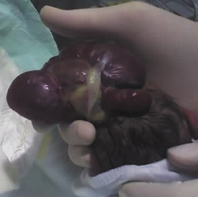
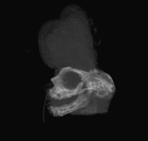
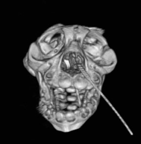
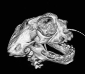
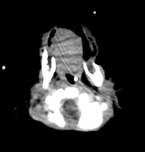
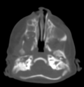
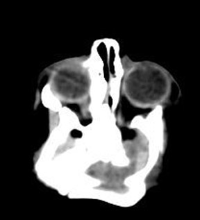
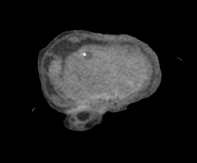
Disorganized brain tissue arising from the base of the cranium. The skull bones are malformed and the posterior cranial fossa is small. The flat bones of the skull are absent.
Discussion
Exencephaly is a congenital malformation along a spectrum that includes acrania and anencephaly (acrania-exencephaly-anencephaly sequence).2,3 In acrania, the flat bones of the skull are at least partially absent, with complete but abnormal development of the cerebral hemispheres. Exencephaly demonstrates a large amount of disorganized brain tissue extending from a malformed skull base (Figure 3).3 Because this brain tissue is covered by only a vascular layer of epithelium, it can be slowly degraded by the amniotic fluid and degenerate into anencephaly.5 The incidence of exencephaly is much lower than that of anencephaly, but the etiology is the same, so the recurrence risk is the same as for other neural tube defects.6 Although the mineralization of the skull is not well established until after 10 weeks, there have been cases of prenatal diagnosis as early as 9 weeks, 3 days proven by postmortem endovaginal ultrasound.2,4 First trimester ultrasound findings of exencephaly include decreased size of the cranial pole compared with the chest, dorsally bulging cranial pole, irregularity of the surface of the cranium2 and echogenic amniotic fluid.3 The etiology of the sequence is not exactly clear, but failed migration of the mesenchyme during the 4th week of development or failed differentiation of the mesenchyme have been suggested.3 Although a significant number of fetuses with open neural tube defects are chromosomally abnormal, this child was chromosomally normal.
Conclusion
The acrania-exencephaly-anencephaly sequence is commonly diagnosed by prenatal ultrasound between 14 and 18 weeks, but some findings may suggest the diagnosis much earlier in gestation. Regardless of gestational age at diagnosis, the prognosis is poor.
Prepared by Dr. Drinkwine, Lieutenant Commander, Medical Corps, United States Navy, and Dr. Holston, Lieutenant, Medical Corps, United States Navy, while at the Naval Medical Center, Portsmouth, VA.
The views expressed in this article are those of the authors and do not necessarily reflect the official policy or position of the Department of the Navy, Department of Defense, or the United States Government.
- Cincore V, Ninios AP, Pavlik J et al. Prenatal diagnosis of acrania associated with amniotic band syndrome. Obstet Gynecol. 2003;102:1176-1178.
- Becker R, Mende B, Stiemer B et al. Sonographic markers of exencephaly at 9 + 3 weeks of gestation. Ultrasound Obstet Gynecol. 2000;16:582-584.
- Bianca S, Ingegnosi C, Auditore S et al. Prenatal and postnatal findings of acrania. Arch Gynecol Obstet. 2005;271:256-258.
- Machado RA, Brizot ML, Carvalho MH et al. Sonographic markers of exencephaly below 10 weeks’ gestation. Prenat Diagn. 2005;25:31-33.
- Chen CP, Chang TY, Lin YH et al. Prenatal sonographic diagnosis of acrania associated with amniotic bands. J Clin Ultrasound. 2004;32:256-260.
- Nawale AJ, Merchant SA, Koteyar SSR et al. Exencephaly: A rare case diagnosed on antenatal ultrasound. Bombay Hospital Journal [serial online]. 2000;42. Available at: http://www.bhj.org/journal/ 2000_4203_jul00/case_520.htm. Accessed June 12, 2006.