Uterine lipoleiomyoma
Images


Prepared by Ashish Chawla, MD, Ajay Thakker, MD, Hemant Telkar, MD, and Nikhil Kamat, MD from the Jupiter Scan Centre, Mumbai, India; and Krantikumar Rathod, MD, Abhijit Raut, MD, and Ashwin Garg, DNB from the Department of Radiology, King Edward Memorial VII Hospital, Parel, Mumbai, India.
CASE SUMMARY
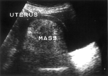
A 65-year-old postmenopausal woman, gravida 2, para 2, presented with leucorrhoea and mild pelvic discomfort. Physical examination revealed a palpable lump arising from her pelvis. Pelvic ultrasound was performed (Figure 1).
DIAGNOSIS
Uterine lipoleiomyoma
IMAGING FINDINGS
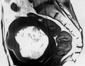
Pelvic ultrasound demonstrated a large, well-defined, hyperechoic mass, measuring 9.9 × 9.9 × 8.9 cm related to the anterior wall of the uterus (Figure 1). Multiple small hypoechoic intramural leiomyomas were also seen. The ovaries could not be identified separate from the mass. In view of these findings, magnetic resonance imaging (MRI) was performed with a 0.5T unit (GE Signa Contour, GE Medical Systems, Milwaukee, WI). MRI showed a large lobulated mass arising from the anterior wall of the uterus, which had hyperintense signals on T1-weighted spin echo (SE) images (repetition time [TR] 420, echo time [TE] 12), which suppressed on a fat-saturation sequence (Figures 2A and 2B). On T2-weighted fast spin-echo (FSE) images (TR 4800, TE 88), the mass appeared inhomogeneous and hyperintense with chemical shift artifact, further suggesting fat content within the mass (Figure 2C).
A subsequent CT scan (GE Dxi/Hi speed) revealed a predominantly fat-containing uterine mass with strands of isodense soft tissue (Figure 3). A preoperative diag-nosis of uterine lipoleiomyoma was made.
PATHOLOGIC FINDINGS
The patient underwent a total abdominal hysterectomy. Gross pathologic examination of the uterus revealed a large, well-circumscribed, solid mass with a yellowish cut surface, mixed with few gray areas. Histopathologic evaluation identified mature adipose tissue and smooth muscle, which are consistent with lipoleiomyomas.
DISCUSSION
A lipoleiomyoma is an uncommon benign uterine neoplasm. To date, only 9 cases have been published in imaging literature. MRI findings have been described in only 4 cases. 1-4 Lipoleiomyomas are composed of mature smooth muscle, fat, and connective tissue. 5 These masses are similar to uterine leiomyomas, in both clinical presentation and course. 6
Lipomatous uterine tumors are typically found in postmenopausal women and are associated with leiomyomas. 5 They usually occur in corpus, predominantly intramurally; however, they may be subserosal. 4,5 A case of ovarian lipoleiomyoma has also been reported. 7 The histologic spectrum includes lipoma, lipoleiomyoma, and fibromyolipoma. 2 The masses may be endophytic or exophytic, with respect to the uterus.
Because fat tissue is not native to the myometrium, various theories have been proposed for the histogenesis of these tumors. A histopathologic study suggested that fatty metamorphosis of smooth muscle cells of a leiomyoma is the most likely etiologic factor in the formation of adipose tissue, rather than fatty degeneration. 8
The differential diagnosis of the lipomatous mass in the pelvis includes: benign cystic ovarian teratoma, malignant degeneration of cystic teratoma, nonteratomatous lipomatous ovarian tumor, benign pelvic lipoma, liposarcoma, 2,7 and lipoblastic lymphadenopathy. 9 Since an asymptomatic lipoleiomyoma requires no treatment and is clinically similar to a leiomyoma, it is important to differentiate these tumors from ovarian teratomas, which require surgical ex-cision. 1 However, associations of lipomatous uterine tumors and endometrial carcinomas with lipoleiomyosarcomas arising in uterine lipoleiomyomas have been reported. 10,11
A uterine lipoleiomyoma usually appears as a well-defined hyperechoic mass encased by a hypoechoic ring on ultrasound. This ring represents a layer of myometrium surrounding the lipomatous mass. 1,2,5,6,12 In the cases of a large mass, as in the present case, ultrasound may not be able to identify the exact organ of origin. CT shows more specific findings, revealing a predominantly fatty mass with areas of nonfat soft-tissue density arising from the uterus. 1,2,5,11 MRI is highly specific, delineating fat tissue as hyper-intense on Tl- and T2-weighted images, with chemical shift artifact. 1,2 Further confirmation of the fatty component may be done using fat-saturation sequences. 2,4 Although contrast-enhanced CT and gadolinium-enhanced MRI findings have been reported in other cases of lipoleiomyomas, 1 these studies were not required in the present case.
Imaging plays an important role in preoperatively identifying the fatty nature and exact intrauterine location of a leiomyoma. Radiologists need to be aware of this entity, which, if large in size, can be mistaken for a more common pelvic mass, such as an ovarian teratoma, on ultrasound. The diagnosis can be confirmed on CT or MRI, which can specifically depict fat content within the tumor.
CONCLUSION
We describe imaging features in a case of a large intramural uterine lipoleiomyoma. Although ultrasound is quite sensitive for the tumor, CT and MRI are more specific. The demonstration of fat in a mass of uterine origin is highly suggestive of a case of lipoleiomyoma. CT and MRI are useful in differentiating uterine lipoleiomyomas from more common fat-containing pelvic masses like ovarian teratomas, which have an altogether different management and prognosis.