Philips CT | Helping Deliver Better Care for More People
In today’s clinical practice, increasing workloads and complexities pose a significant challenge to the delivery of high-quality imaging. Philips CT systems are designed to meet these demands with meaningful innovation at their core. By integrating advanced AI technologies and intelligent workflows, they empower healthcare providers worldwide to elevate the standard of care while addressing the patient and cost pressures that imaging organizations face today.
Spectral CT 7500: Lead with Spectral Speed
As a large teaching hospital with one of the largest oncology clinics in Europe, the University Hospital of Cologne in Germany performs about 220,000 imaging studies a year. This includes a wide range of cases, from cardiac and neurology, to body, MSK and oncology. In 2022, the facility installed the Philips Spectral CT 7500 in the emergency department.
With detector-based spectral CT always on, radiologists have certainty in routine spectral imaging, without an increase in radiation dose. The detector-based spectral CT system delivers rich spectral data in a single scan, on demand, even retrospectively, all while maintaining a simple and efficient process. Fast scanning and reconstruction speed up time to diagnosis, which is critical in the ED where CT serves as the primary imaging tool in high-pressure situations. Additionally, results are immediately accessible in any reading environment via Philips Spectral Magic Glass on PACS.
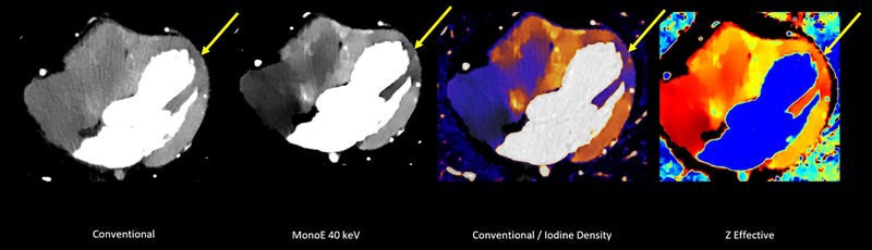
“Now dual-energy data is collected all the time. While it can be integrated into a full conventional image data set, you can also get spectral results after just a couple minutes of reconstruction time,” said Nils Grosse Hokamp, MD, PhD, MBA, EDiR, Associate Professor of Radiology and Experimental Radiology and Section Lead Urogenital Radiology in the Institute for Diagnostic and Interventional Radiology at the University Hospital of Cologne.
This detector-based spectral CT system offers spectral results 100% of the time for excellent image quality, low dose and easy workflow, with no special protocols or separate reconstructions.
“The virtual monoenergetic images are better than conventional image. I read those images first, and I don't worry about the conventional images anymore. Overall, the Spectral CT 7500 delivers better images in both routine daily workflow and characterization of findings,” said Dr. Grosse Hokamp.
The Spectral CT 7500 provides advanced diagnostic capabilities with a single spectral scan, transforming patient care by enabling accurate diagnoses, expediting treatment and reducing the need for invasive procedures. It allows the coronaries and myocardium to be assessed in a single exam, enabling appropriate patients to avoid an invasive and expensive procedure in the cath lab, while supporting treatment with minimal delay for ED and trauma patients.
Workflow is easy. Staff can scan in seconds for conventional CT with immediate access to spectral CT results, helping eliminate the need for multiple CT scans or additional scans from other modalities. This allows the facility to avoid repeat scans, which directly impacts the bottom line. For example, when imaging an adrenal mass, spectral results can differentiate a benign adenoma from a malignant tumor, which avoids the time and costs associated with an additional MRI exam.
As healthcare becomes increasingly complex, the Spectral CT 7500 simplifies the patient care journey at the University Hospital of Cologne by reducing uncertainty and saving valuable time, scans and costs.
Now radiologists can perform fast, low-dose scans without compromising speed, power or field of view. “It doesn't matter where we position patients because we have such a wide field of view in the Z direction that we can get good images in less time,” said Dr. Grosse Hokamp.
Spectral 7500 requires radiologists to adapt to a new imaging approach, transitioning towards more functional imaging in CT, traditionally known for its anatomical insights. While it introduces a new learning curve for radiologists, Dr. Grosse Hokamp says it’s worth the benefits. “The 7500 is a next-generation spectral CT system. We have a high acquisition speed with spectral results, allowing us to image a wide range of body structures with precision and efficiency. Spectral is a great thing to have and, for me, it's the only way that CT should be,” said Dr. Grosse Hokamp.
Confidently assess myocardial hypoperfusion with spectral.
CT 5300: Next-Level Confidence, Empowered Workflow and Value For A Lifetime
Foundation IRCCS San Gerardo dei Tintori in Monza, Italy, performs approximately 200,000 imaging studies every year. As the largest trauma centers in northern Italy, the hospital sees many emergency cases, which is why it recently installed the Philips CT 5300 in the Emergency Department (ED). This speeds up the diagnostic capacity in emergency situations, particularly in “time-dependent” cases such as strokes and hemorrhages, so patients can be quickly directed to the most appropriate treatment pathway.
“It delivers real-time images, so we immediately see the results and don’t have to wait. With this scanner, we’re able to address the needs of patients in all areas of the hospital,” said Davide Ippolito, MD, a diagnostic radiologist at Foundation IRCCS San Gerardo dei Tintori and a professor of radiology at the University of Milan.
The Philips CT 5300 leverages artificial intelligence (AI) for new clinical capabilities and workflow advances. It includes CT Smart Workflow, a comprehensive suite of AI-enabled capabilities that delivers precision in dose, speed, and image quality for a wide range of studies. These include oncology, neurological, cardiac, pediatric, and trauma studies.
With Precise Position1, an AI-enabled smart camera saves time while improving accuracy of vertical centering relative to manual positioning by up to 50% during scan prep2. It reduces patient positioning time by up to 23% and increases consistency from user to user by up to 70%.2
“Precise Position increases efficiency because the protocol is more reproducible. AI helps us correctly center the patient at the isocenter with a camera that recognizes anatomical landmarks, which makes results consistent for follow-up imaging, and we can reduce patient preparation time,” said Dr. Ippolito
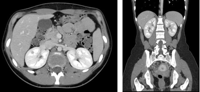
Low dose abdominal scan reconstructed with Precise Image. Scan parameters: 100 kVp, 99 mAs, CTDIvol 4,82 mGy, DLP 250mGy*cm.
The powerful Precise Image AI reconstruction delivers 80% lower radiation dose, 85% lower noise and 60% improved low-contrast detectability.3 With Precise Image, Foundation IRCCS San Gerardo dei Tintori reduced dose in body imaging scans tube voltage from 120 kVp to 100 kVp and, in cardiac imaging, from 100 kVp to 80 kVp – without an impact on image quality. “We were able to reduce dose anywhere from 33% to 80%, depending on anatomy. Even better, image quality is the same as those taken at our highest dose. That's a great gain from a clinical point of view and for patient safety,” said Dr. Ippolito.
This simultaneously delivers cost savings and drives sustainability measures. Plus, the CT 5300 delivers value for a lifetime through remote services with predictive monitoring and Philips’ innovative Tube for Life guarantee.4
Both Dr. Ippolito and Dr. Grosse Hokamp agree that these next-generation CT scanners are delivering deeper clinical insights and helping address more complex cases. By integrating advanced AI capabilities and intelligent workflows, these systems deliver precision, speed and image quality that empower healthcare providers to address patient needs with confidence and efficiency. From reducing radiation dose and energy consumption to improving diagnostic accuracy and sustainability, these innovations are setting new standards for care.
References
- Patients below the age of 18 are not supported.
- Based on Philips in-house assessment by five clinical experts, comparing manual positioning versus Precise Position in 40 clinical cases using a human body phantom.
- In clinical practice, the use of Precise Image may reduce CT patient dose depending on the clinical task, patient size, and anatomical location. A consultation with a radiologist and a physicist should be made to determine the appropriate dose to obtain diagnostic image quality for the particular clinical task. Dose reduction assessments were performed using reference body protocols with 1.0 mm slices at the “Smoother” setting, and tested on the MITA CT IQ Phantom (CCT189, The Phantom Laboratory) assessing the 10mm pin and compared to filtered-back projection. A range is seen across the 4 pins, using a channelized hoteling observer tool, that includes lower image noise by 85% and improved low-contrast detectability from 0% to 60% at 50% to 80% dose reduction. NPS curve shift is used to evaluate image appearance, as measured on a 20 cm water phantom in the center 50 mm x 50 mm region of interest, with an average shift of 6% or less.
- Life of the product is defined by Philips as 10 years. Tube for Life guarantee availability varies by country. Please contact your local Philips sales representative for details.