IQon Spectral CT: Improving Patient Management in Neurology
Images


Supplement to November-December 2019 Applied Radiology. Sponsored by Philips Healthcare.
The radiology team at Einstein Medical Center in Philadelphia, PA, is changing the way it helps fight stroke. A key weapon in their arsenal: Philips’ IQon Spectral CT.
Stroke is a leading cause of death and disability worldwide; imaging plays a critical role in evaluating suspected cases of the condition in patients who present to the emergency department, as well as in assessing the results of treatment.
As part of their comprehensive imaging protocol, radiologists at the flagship hospital of the namesake Einstein Health Network (Einstein) are leveraging IQon Spectral CT technology to image ischemic stroke patients before and after recanalization therapies. And along with recent changes they have made to their clinical workflow, they’re reducing costs and improving care and patient experience.
Optimal imaging
Given the complexity of stroke and its effects, optimizing imaging protocols is critical, especially with respect to selecting the right modality and the ideal acquisition technique from among many available such techniques, needed to generate the best possible patient outcome.
Einstein’s radiology department uses Philips’ IQon Spectral CT as its technology of choice in stroke imaging. Ryan Lee, MD, Einstein Radiology’s Vice Chair of Quality and Safety and Section Chief of Neuroradiology, says spectral CT has made a significant impact on stroke management at the medical center. In fact, longstanding protocols were recently changed to require that all post-interventional scans be acquired on the IQon Spectral CT.
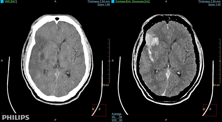
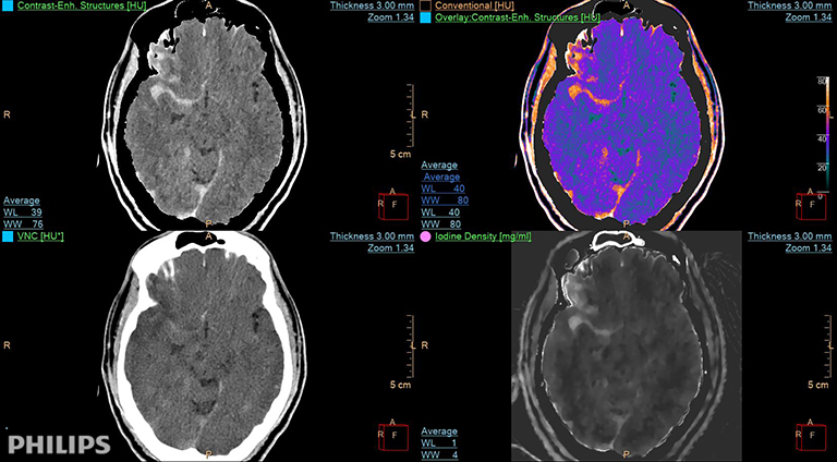
It’s common, Dr. Lee says, to see density in the infarct bed on the brain CT scans immediately obtained after intervention.
“Typically, this is contrast staining, but it could be hemorrhage,” he says, explaining that a hemorrhage finding warrants more intense follow-up.
“We’ve found that, using IQon Spectral CT, we can more definitively differentiate between contrast staining and blood in these images,” Dr. Lee says. “Because I have spectral data, I can manipulate the images to get specific reconstructions to separate blood from contrast. It didn’t take very long for me to see the potential to eliminate the follow-up CT for confirmation.”
Change for good
Indeed, Dr. Lee proposed permanently eliminating confirmation CT scans of post-stroke treatment patients based on the results provided by the IQon Spectral CT, and both the neurology and interventional radiology teams agreed. This simple change to the workflow has improved staff and patient experience and reduced costs by doing away with unnecessary exams. The neurology and interventional radiology physicians are pleased with the change, he says.
“The main benefit is the increased confidence we have that it is contrast staining and not hemorrhage, allowing us to not obtain another follow-up head CT. As a result, we are improving our overall quality of care with this protocol change,“ says Dr. Lee, adding that by eliminating these additional scans, “We’re relieving some of the anxiety that comes from waiting for the ‘all clear’ after the interventional procedure.”
Routinely better
Having worked with the IQon Spectral CT for about a year now, Dr. Lee says the benefits of spectral technology are also apparent during image reconstructions. IQon Spectral CT reconstructions are completed on the scanner; then Dr. Lee performs advanced analysis using IntelliSpace Portal, allowing him to obtain a comprehensive understanding of the patient’s neurovascular status. When complete, the images are saved to the medical center’s picture archiving and communications systems (PACS).
“On the post-processing side, there was a bit of a learning curve in understanding the different reconstructions available, but it has become second nature to those of us that routinely use it,” notes Dr. Lee, who adds that the workflow change has had little impact on scheduling or accessibility. The IQon is located in Einstein’s emergency department, which also gives that department’s physicians easy access when necessary for cases of suspected stroke.
Sharing experience
Dr. Lee says there are plans to formalize, write, and publish the workflow data and resulting changes in stroke patient management so that other institutions can benefit from Einstein’s experience. Additionally, the radiology team is making the most of the experience by adapting the same changes to other clinical areas and procedures as appropriate, such as in middle meningeal artery embolization, which is used to reduce the size of patients’ chronic subdural hematoma.
“Middle meningeal artery embolization, says Dr. Lee, “can result in a similar diagnostic dilemma as those for post-intervention stroke patients. It is difficult to discern contrast from hemorrhage on our regular scanners just like with the post-intervention stroke cases, so we are performing these studies on the IQon scanners as well.”
Citation
IQon Spectral CT: Improving Patient Management in Neurology. Appl Radiol.
November 13, 2019