Clinical impact of diffusion tensor imaging
Images


Dr. Tanenbaum is Section Chief of MRI, CT, and Neuroradiology, New Jersey Neuroscience Institute and Edison Imaging-JFK Medical Center, Edison, NJ; and Assistant Professor, Department of Neuroscience, Seton Hall School of Graduate Medical Education, South Orange, NJ. He is also a member of the editorial board of this journal.
Selected images and content from this article originally appeared in the January 2006 issue of Diagnostic Imaging magazine and are reprinted with permission. Adapted and reprinted from: Tanenbaum LN. Diffusion tensor imaging delivers crucial information: Use in clinical practice to work up on all lesions considered for surgical resection can reduce impact on eloquent structures. Diagn Imaging (San Franc). 2006;28(1):39-45.
Diffusion magnetic resonance (MR) imaging became widely available for clinical use in the late 1980s; it revolutionized the MR imaging of patients with suspected brain infarction and had an enormous impact on the characterization of focal brain lesions. Recent advances in MR have enabled diffusion tensor imaging (DTI), offering new insights into the nature and integrity of ordered brain white matter (WM) pathways. Imaging brain anisotropy can yield useful information about WM integrity and exhibit pathology that is occult to conventional imaging techniques. Anisotropy imaging can also provide information about ordered WM tracts, such as directional orientation and connectivity (tractography), which can be critical in surgical planning and useful in the understanding of certain developmental and acquired disease states.
Physical principles
By manipulating magnetic field gradients, MR images can be sensitized to diffusion--the random, thermally driven motion of water molecules in tissue. Diffusion is anisotropic (directionally oriented) in WM tracts, as axonal membranes and myelin sheaths present barriers to water motion in directions other than parallel to fiber orientation. The direction of maximum diffusivity coincides with WM fiber tract orientation. Water movement is essentially isotropic and random in brain gray matter.
This directional information is contained in the diffusion tensor, a mathematical model of diffusion in a 3-dimensional (3D) space. The tensor is a matrix of numbers derived from diffusion measurements in multiple directions (at least 6) from which diffusivity in any direction can be estimated and the direction of maximum diffusivity can be determined. The tensor matrix can be visualized as an ellipsoid. The diameter in any direction estimates the diffusivity in that direction, and the major principle axis is oriented in the direction of maximum diffusivity.
The degree to which the diffusion tensor shape differs from that of a sphere (random, isotropic motion) represents anisotropy (ordered motion). With DTI, the degree of anisotropy and the orientation of fibers can be mapped, which provides an opportunity to study WM architecture and to evaluate fiber integrity.
Data analysis and display
Two-dimensional (2D) and 3D tract rendering is accomplished with commercially available software that typically resides in a scanner console or a free-standing workstation. Two-dimensional techniques include magnitude rendering of WM anisotropy and color encoding of tract orientation. With 3D techniques, the ordered WM tracts can be displayed in space against the background of the routine structural information provided by MR imaging-a technique useful in preoperative neurosurgical assessment. While individual tract parsing is imperfect-in some circumstances incompletely rendering individual tracts, and in others insufficiently differentiating and extracting adjacent tracts-these techniques are still clinically efficacious.
White matter fiber classification
White matter fiber tracts are classified as association , projection , or commissural fibers. Association fibers interconnect cortical areas in each hemisphere. Projection fibers interconnect cortical areas with deep nuclei, the brain stem, the cerebellum, and the spinal cord. Commissural fibers interconnect similar cortical areas between opposite hemispheres.
Clinical utility
Parameters derived from the diffusion tensor, such as relative and fractional anisotropy, have been used to evaluate multiple disease states, including adrenoleukodystrophy, multiple sclerosis, and acquired immune deficiency syndrome. Anisotropy imaging has been studied in the evaluation of hypertensive encephalopathy, leukoariosis, and aging changes.
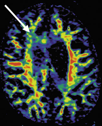
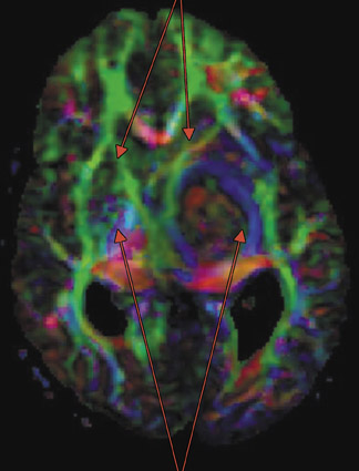
Use in neuropsychiatric disorders, such as traumatic brain injury, reveals reductions in anisotropy corresponding to regions of injured brain. Schizophrenic patients show a reduction in anisotropy in frontal WM pathways similar to the pattern of reduction that is seen with perfusion imaging (Figure 1).
In Alzheimer's disease, reduced anisotropy has been reported in the corpus callosum as well as in the fronto-occipital and thalamofrontal tracts, which corresponds to the loss of coherence between the frontal and occipital lobes that is seen on electroencephalographic testing. 1
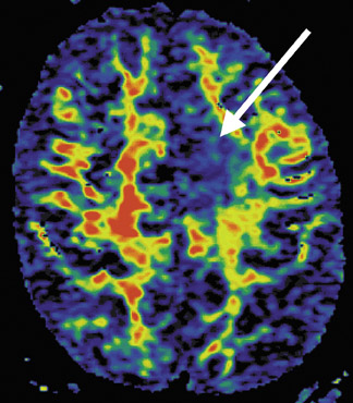
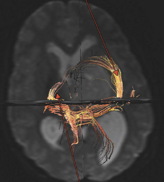
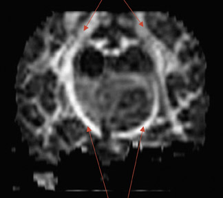
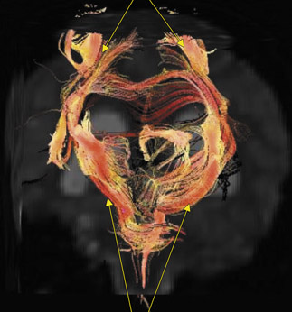
Anisotropy imaging and DTI have been utilized in the workup of patients with epilepsy, revealing a loss of anisotropy in tracts such as the fornix and hippocampal stria (Figure 2). This supplements the information obtained with structural MR imaging and electroencephalographic testing and assists in the localization of atrophic lesions such as hippocampal sclerosis.
Diffusion tensor imaging has been used to investigate brain development and to assist in understanding the organization of the brain WM in developmental brain abnormalities, often demonstrating additional findings beyond those that are seen with conventional MR imaging. 2
Surgical planning
The preservation of vital cerebral function while maximizing lesion resection is the principal goal in brain neurosurgery. Cortical mapping can be accomplished intraoperatively with electrocortical stimulation. Preoperatively, functional MR imaging (fMRI) techniques (blood-oxygenation-level-dependent [BOLD] imaging) are used in the localization of eloquent cerebral cortex. Neither technique provides information about WM tracts in or adjacent to brain lesions. Two-dimensional and 3D WM tractography techniques can be very powerful in elucidating relationships of deep brain lesions to eloquent brain structures, assisting in estimation of the impact of surgical intervention and lesion resection on brain function. 3
At the New Jersey Neuroscience Institute, DTI is part of the imaging workup of all lesions being considered for surgical resection. White matter imaging is used to estimate the relationship of the lesion to tracts responsible for brain activity, such as motor function (corticospinal tracts). Diffusion tensor imaging techniques are also used to estimate the effect of surgical intervention on residual language (superior longitudinal [arcuate] fasciculus) or vision function (geniculocalcarine [optic radiations] tracts). Diffusion tensor tractography, which requires approximately 5 minutes to scan and is easily processed with commercially available software, is practical and easily integrated into the armamentarium of techniques of the high-end neuro-oriented clinical practice. 4
Case study I
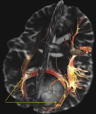
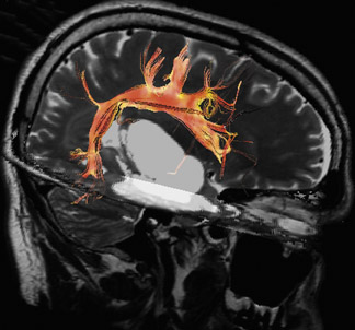
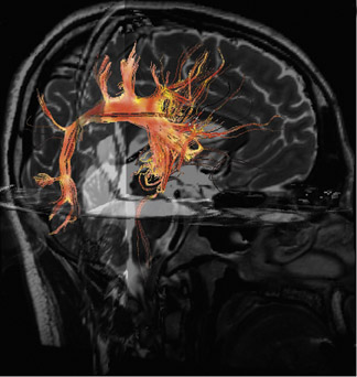
A 13-year-old girl, diagnosed at an outside institution with a low-grade thalamic glioma, was referred to the New Jersey Neuroscience Institute for evaluation for resection. The treating neurosurgeon requested a 3T MR study for detailed anatomic delineation with DTI and diffusion tensor tractography (DTT) for functional localization. In many cases, the shortest approach to a tumor may not be the safest, as eloquent WM pathways may stand between the surface of the brain and the lesion. The combination of high-resolution 3D structural and DT functional information can be critical in planning the least destructive path to a lesion.
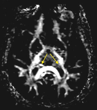
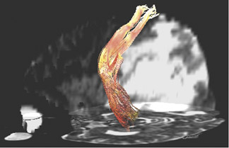
Imaging revealed a well-circumscribed, nonenhancing left thalamic mass distorting the local anatomy and obscuring relationships with the adjacent internal capsule. Definitive localization of the blue corticospinal fibers coursing in a cephalocaudal direction was made possible by the directionally encoded 2D tensor images (Figures 3 and 4).
These images, along with 3D tractograms seeded and grown from the ipsilateral precentral gyrus WM, showed that the lesion was displacing the posterior limb of the internal capsule laterally and inferiorly. The anterior limb of the internal capsule, rendered in green as its fibers course anteroposteriorly, was displaced anteromedially. Fibers that run right to left are depicted in red by convention. Armed with this functional information, the surgeon resected from a medial approach. The surgery went well, and the patient was discharged several days later. She had no motor deficit after resection.
Case study II
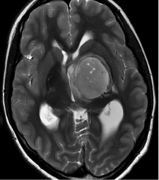
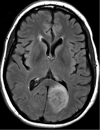
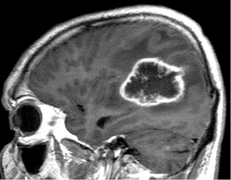
A 35-year-old man presented with seizures. 3T MR imaging revealed a low-grade glioma within the occipital lobe (Figure 5). The goal of neurosurgery was to resect the lesion with minimal disruption of visual function. Tractography revealed that the lesion was medial to the optic radiations as well as medial and inferior to the superior longitudinal fasciculus, which led to a paramedian posterior approach to lesion removal and minimal impact on visual and language function.
Case study III
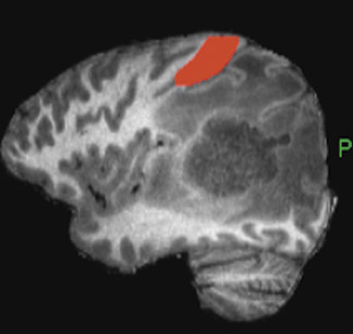
A 30-year-old woman with a low-grade temporal lobe glioma was being considered for surgery. There was significant concern over the possibility of reduced language function after resection. Diffusion tensor imaging fused to a high-resolution 3D fast spin-echo anatomical study showed the superior longitudinal (and arcuate) fasciculus separate from the lesion and indicated the best approach to resection (Figure 6).
Case study IV
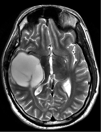
A 40-year-old patient with a recurrent high-grade glioma was referred for consideration of lesion debulking. Blood-oxygenation-level-dependent imaging readily marked the central sulcus and showed that the anterior border of the lesion was posterior to the eloquent cortex at the brain surface (Figure 7). The neurosurgical plan was to debulk the lesion by removing the enhancing core deep within the brain. The localization of function with respect to the deeper portions of this large lesion was facilitated by 3D DTT of the WM fibers. The corticospinal fibers were clearly displaced and bowed anteriorly by the tumor. Lesion debulking did not produce a motor deficit. Diffusion tensor imaging is a critical adjunct to BOLD imaging in the routine assessment of patients who are being considered for neurological surgery.
Conclusion
Advanced imaging tools using DTI and DTT are making a significant impact in the clinical imaging of patients with neurological disease. Yielding structural and functional information about ordered WM pathways in the brain, DTI/DTT assists in the understanding of various disease states, identifying conditions occult to structural imaging and providing relational information that is critical to neurosurgical decision making. The studies, which can be acquired and processed in a practical and efficient manner, are applicable in any high-level neuroimaging practice setting.
Related Articles
Citation
Clinical impact of diffusion tensor imaging. Appl Radiol.
January 22, 2007