3D Whole Breast Ultrasound Tomography System Receives FDA Approval
 The US Food and Drug Administration (FDA) has granted premarket approval (PMA) of SoftVue 3D Whole Breast Ultrasound Tomography System from Delphinus Medical Technologies for use as an adjunct to digital mammography in the screening of asymptomatic women with dense breast tissue.
Clinical evidence has demonstrated that SoftVue enhances dense breast screening and identifies up to 20% more cancers with greater accuracy and potentially fewer biopsies than full field digital mammography (FFDM) alone. The SoftVue exam is completed with no compression or radiation, and the PMA indication for use allows SoftVue exams to be performed at the same appointment as screening mammograms, facilitating a streamlined workflow and rapid delivery of results.
“SoftVue plus mammography finds more cancers when screening women with dense breasts. The FDA clearance for same day imaging optimizes workflow while providing unparalleled patient comfort with no compression or radiation, will make patient screening more comprehensive, efficient and effective. It is exciting to have a new cutting-edge technology that detects more cancers and does so with fewer biopsies,” said Dr. Rachel Brem, director for breast imaging at George Washington University Hospital, Washington DC and a member of Delphinus’s board of directors.
SoftVue was developed to address the unmet clinical needs for early breast cancer detection in individuals with dense breast tissue (fibrous tissue that can hide cancerous lesions on mammogram) and provides a new annual screening solution for this population. The system identifies more cancers, with fewer callbacks, using a proprietary TriAD (Triple Acoustic Detection) technology that effectively characterizes tissue by recording reflection, speed and direction of sound waves moving through breast tissue, unlike traditional ultrasound which utilizes only reflection.
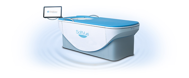
During the exam, the patient relaxes on her stomach with her breast submerged in a warm water bath. The breast is comfortably stabilized and centered with a disposable Sequr Breast Interface gel pad. Imaging is performed with a proprietary 360-degree ring transducer, scanning each breast from chest wall to nipple in an average of three minutes, capturing new images every two millimeters. The captured signals are then analyzed using sophisticated algorithms that provide cross-sectional slices of the entire volume of breast tissue.
“Our SoftVue System delivers a breakthrough in tissue characterization and improves the ability to find cancers in dense breast patients. It will be a game changer that will transform clinical practice with a fundamentally new, and highly impactful approach,” said Mark J. Forchette, president and chief executive officer at Delphinus. “The SoftVue PMA approval opens the door to a technological advance in dense breast screening that will help physicians save lives.”
Delphinus executed a Prospective Case Collection (PCC) study that served as the image database for the cancer and non-cancer cases that were subsequently used in a series of multi-reader, multi-case (MRMC) studies conducted by the University of Chicago. The PCC initiative began in the summer of 2017 and ultimately enrolled more than 8,500 asymptomatic patients in c or d density categories at 10 leading breast imaging centers around the US. A team of radiologists analyzed more than three million coronal plane images generated by the initiative.
The pivotal MRMC demonstrated that SoftVue detects and distinguishes between normal (BI-RADS 1 and 2) and abnormal (BI-RADS 3 and 4) lesions with an increase in both sensitivity of 20% and specificity of 8%, as compared to FFDM alone. Additionally, 95% of patients surveyed after their SoftVue exam indicated that they would recommend SoftVue to other women.
“With the FDA approval of SoftVue, we now have a critical new imaging tool for screening women with dense breasts to enable the earlier detection of breast cancer,” said Dr. Mary Yamashita, lead investigator of the SoftVue PCC clinical trial and Clinical Associate Professor of Radiology at Keck School of Medicine, University of Southern California, Los Angeles. “We have immense gratitude for the patients and their families, clinicians and technologists that worked so hard to make this rigorous study a success. We have expanded the tools available for breast cancer diagnosis, particularly in a population of women most in need of this technology.”
Related Articles
Citation
. 3D Whole Breast Ultrasound Tomography System Receives FDA Approval . Appl Radiol.
October 13, 2021