Using Explainable AI For Medical Image Processing and Cancer Screening

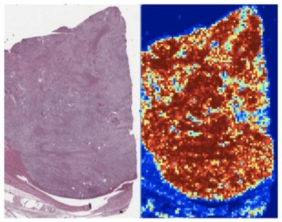
Kidney renal clear cell carcinoma

Example of WSI kidney tissue
AI explainability is a part of AI functionality that is responsible for explaining how the AI came up with a specific output. MobiDev experts are now using explainable AI for medical image processing and cancer screening.
Computer vision techniques are actively used in the processing of medical images, such as Computed Tomography (CT), Magnetic Resonance Imaging (MRI), and Whole-Slide images (WSI). For example, WSI of human tissues can reach 21,500 × 21,500 pixel size and even more. However, since WSI is a large image, it takes significant amounts of time, attention and qualification to analyze. Traditional computer vision methods also require too many computational resources for end-to-end processing.
In terms of WSI analysis, explainable AI can act as a support system that scans image sectors and highlights regions of interest with suspicious cellular structures. The machine won't make any decision, but will speed up the process and make the work of a doctor easier. This is possible because WSI is more accurate and quicker in terms of image scanning, giving less chance to omit specific regions. AI support system highlights the regions of high risk where cancer cells are more likely to be. This eliminates the need to physically analyze the whole image of a kidney, providing hints for medical expertise and attention.
Citation
. Using Explainable AI For Medical Image Processing and Cancer Screening. Appl Radiol.
July 27, 2022