Sino-orbital pathologies: An approach to diagnosis and identifying complications
Images
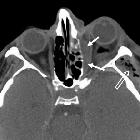
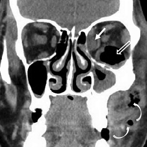
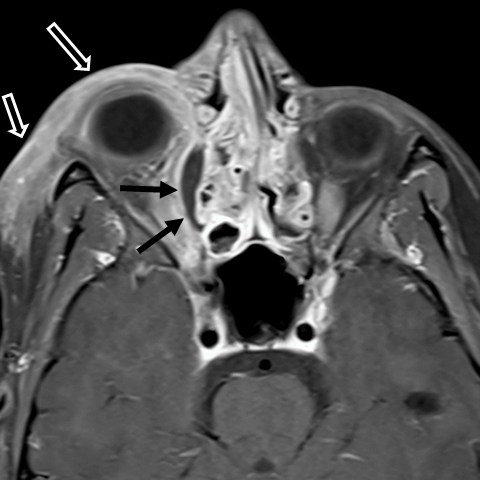

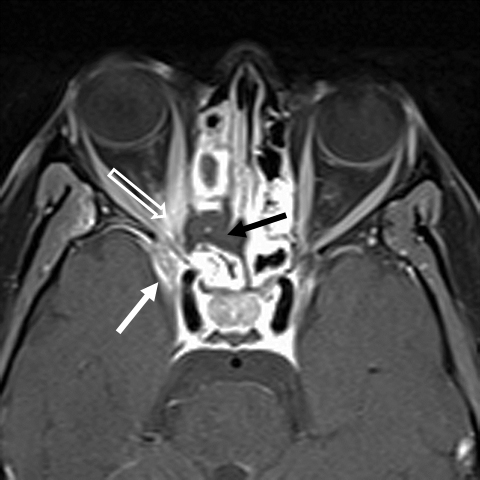


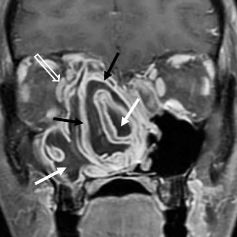

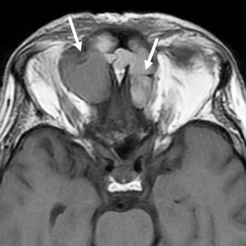
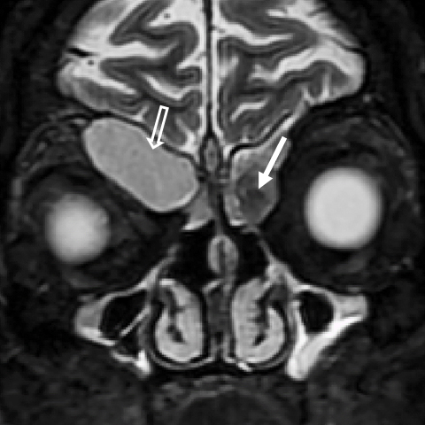
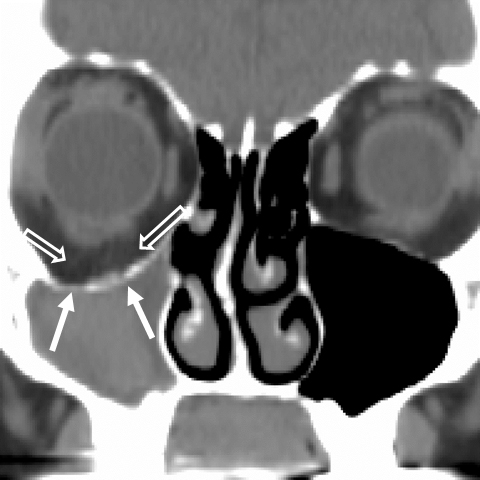
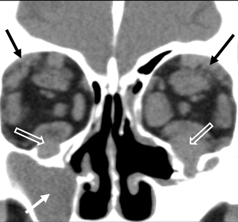


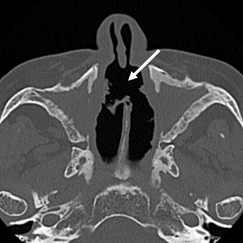

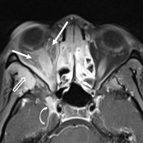
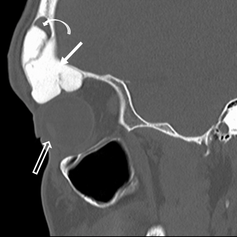

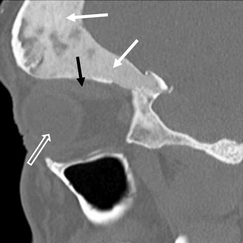
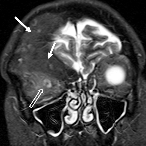
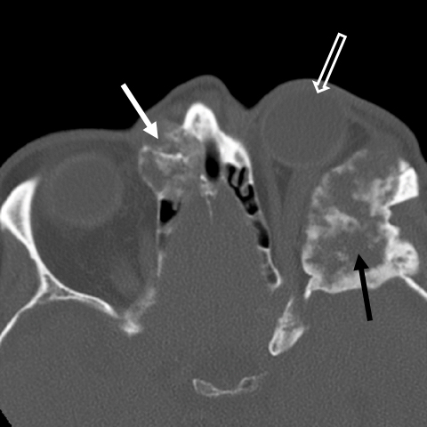
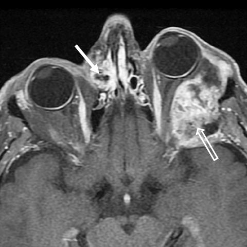

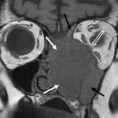
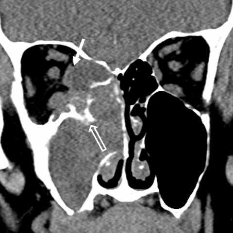
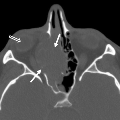
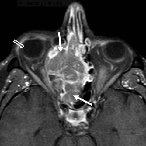
A variety of diseases are unique in their ability to involve both the sinonasal (SN) cavities and the orbits. It is more common for SN pathology to affect the orbit than the reverse, and primary sinus pathology may initially present with predominantly orbital, rather than sinus, symptomatology. Imaging can be very helpful for localizing the site of origin of pathology, for narrowing the differential diagnosis, and for identifying additional findings that may impact medical or surgical management.
We present a practical approach to diagnosing sino-orbital pathologies with an emphasis on distinguishing imaging features of lesions and recognizing impact on important adjacent anatomic structures. Although we cannot demonstrate all of the lesions that can affect both compartments, our hope is that you will be able to diagnose the cases we describe most of the time.
Lesion classification
Disease entities affecting the sino-orbital region may arise primarily in the SN cavities, the orbits, or the surrounding bones; or they may result from secondary involvement by systemic disorders. The incidence of orbital involvement by SN disease is much more common than sinus involvement by orbital disease. We will discuss a variety of pathologies that may involve both compartments and have classified these sino-orbital pathologies broadly into four groups: 1) Infectious and inflammatory conditions; 2) Granulomatous disease; 3) Fibro-osseous lesions; and 4) Neoplasms.
Infectious and inflammatory conditions
Infectious and inflammatory conditions of the sino-orbital region usually arise in the SN cavities and spread to the orbit. Conditions that present acutely include cellulitis, subperiosteal phlegmon/abscess, and acute invasive fungal rhinosinusitis (AIFRS). Chronic inflammatory conditions include allergic fungal sinusitis, mucocele, IgG4-related disease, and acquired maxillary atelectasis. These entities affect the orbit either by direct extension or by distortion of the orbital walls.
Acute bacterial sinusitis
Orbital complications appear most often in frontal or ethmoid sinusitis and are most common in children.1 In adults, orbital complications are more common in immunocompromised and diabetic patients. Symptoms of orbital involvement include an erythematous swollen eye, proptosis, and impaired ocular motility. Most cases are treated non-surgically; however, immediate surgical intervention is recommended in patients with vision loss as an initial symptom, in patients with rapid vision loss over time, and in those without a response to 48 hours of intravenous antibiotic therapy.2
Infection may spread directly into the orbit trans-osseously, hematogenously, or by retrograde extension via the valve-less diploic veins.1 The thin lamina papyracea does not offer much resistance to the spread of infection from the ethmoid sinuses. The periorbita is more resistant and fluid/pus may accumulate between the bone and the periosteum. Subperiosteal phlegmon/abscess is the most common imaging finding in sinogenic orbital infection.3 Of note, orbital and intracranial extension may coexist and early recognition is critical.
On imaging, orbital fat stranding is an early sign of periorbita breech.3 Phlegmon can be difficult to distinguish from frank abscess (Figure 1). Orbital abscess typically appears as a rim-enhancing, fluid attenuation (CT) or signal (MRI) collection along the medial wall or roof of the orbit (Figure 2).
Acute invasive fungal rhinosinusitis
Acute invasive fungal rhinosinusitis (AIFRS) is a rapidly progressing infection with high mortality, reportedly 50-80%.4 The initial presentation may be nonspecific, with nasal discharge or fever; however, visual symptoms and neurological deficits may rapidly develop. AIFRS occurs almost exclusively in two groups of patients: immunocompromised patients, particularly individuals with cellular immune deficiency such as HIV/AIDS; and poorly controlled diabetics.4 Fungal infection in the diabetic group is most often caused by organisms in the Zygomycetes order such as Mucor. 4 In the immunocompromised group, Aspergillus species are responsible for up to 80% of AIFRS cases. 4 The infection typically starts in the nasal cavity with ulceration of the turbinates and nasal septum. Infection then spreads to the paranasal sinuses, then to the orbit and cranial cavity.
Successful treatment requires prompt diagnosis and it is often our duty as radiologists to identify early findings on imaging. MRI is superior to CT in evaluating orbital and intracranial extension. CT and MR angiography may help to identify vascular narrowing/occlusion, particularly of the cavernous segments of the internal carotid arteries in cases of sphenoid fungal disease.
On imaging, early disease may appear as mucosal thickening or soft tissue in the SN cavities with a predilection for the ethmoid and sphenoid sinuses. 4 Ethmoid disease can spread easily into the medial orbit and sphenoid disease, into the orbital apex. On CT, early orbital involvement shows infiltration and soft tissue stranding in the orbital fat surrounding the extraocular muscles (Figure 3A). Bone erosion may or may not be present, as the organisms are angioinvasive and infection can spread along the blood vessels traversing the bone. On MR, the presence of areas of hypointense long TR signal related to the presence of fungal hyphae and metal chelates may be seen. Areas of tissue necrosis, with absence of enhancement, are characteristic of advanced disease (Figure 3B). AIFRS can also appear mass-like and should not be mistaken for neoplasm.
Allergic fungal sinusitis
Allergic fungal sinusitis (AFS) is the most common form of fungal sinusitis, is noninvasive and unrelated to invasive fungal pathology.5 AFS is common in warm, humid climates and occurs in young, immunocompetent patients, often with a history of atopy, including allergic rhinitis and asthma. AFS is characterized by the presence of thick, eosinophilic fungal-laden mucin that has been described as looking like peanut butter. AFS typically involves multiple sinuses with opacification and expansion of the involved sinuses.4 AFS involvement of the ethmoid sinuses is most likely to cause orbital symptomatology, including proptosis, diplopia, and epiphora. Orbital extension is limited by the periorbita and can usually be treated endoscopically.
The CT imaging features of AFS are characteristic: The sinuses are expanded with material that is hyperdense centrally with a peripheral rim of low attenuation (Figure 4). The signal intensity of the allergic mucin varies depending upon the water and protein content. Profound low T2 signal may mimic air (Figure 5A). The hypointense long TR signal was initially thought to be related to hemosiderin accumulation in the mucin, but now is believed to be related to deposition of heavy metals such as iron, magnesium, and manganese concentrated by the fungal organisms.5,6 Correlating the long TR sequences with T1 imaging and/or CT is important to confirm the degree of opacification. Inflamed mucosa at the periphery enhances with contrast, but there is no central enhancement; this helps to differentiate AFS from neoplasm (Figure 5B). Polyposis often occurs with AFS; excluding the presence of polyps in severe cases can be difficult.
Mucocele
Mucoceles result from sinus obstruction with subsequent expansion and are most common in the frontal and anterior ethmoid sinuses. Patients may present with proptosis, diplopia, and inferior (frontal) or lateral (ethmoid) globe displacement. Mucocele formation in the posterior ethmoid and sphenoid sinuses or in variant air cells (sphenoethmoidal/“Onodi” cells) are uncommon, but are more likely to cause optic neuropathy and cranial nerve palsies due to proximity to the optic nerve and cavernous sinuses. Devastating vision loss may rapidly occur if mucoceles in these variants cells become infected (mucopyocele).7
Diagnosing a mucocele on CT or MRI is usually not challenging, as the imaging features are fairly characteristic. CT reveals smooth expansion of the walls of the sinus with or without foci of bone dehiscence (Figure 6). On MR, the contents typically show low T1 and high long TR signal with only minimal peripheral enhancement. The attenuation (CT) and signal (MR) characteristics of mucoceles vary with the chronicity and protein content of the lesion (Figure 7). The presence of a thick rim of enhancement, fluid-fluid level, and/or surrounding soft tissue infiltration suggests mucopyocele formation.
Acquired maxillary sinus atelectasis
Acquired maxillary atelectasis, also known as “silent sinus syndrome,” affects the orbit by an ‘ex vacuo” mechanism, increasing the volume of the orbit. This entity may go unrecognized and is likely underdiagnosed.8 The typical patient presents with painless, progressive enophthalmos and hypoglobus. Although symptoms of chronic sinus disease often are not present, chronic maxillary sinus obstruction leads to negative pressure in the antrum, gradual inward retraction of the sinus walls, including the orbital floor, increased orbital volume, and enophthalmos.8
Imaging shows opacification and volume loss in the affected maxillary antrum, inward retraction of the sinus walls, obstruction of the maxillary infundibulum, lateralization of the uncinate process, widening of the retroantral fat pad, and widening of the ipsilateral middle meatus (Figure 8). The maxillary walls may be thinned, normal or slightly thickened. Anterior bowing of the posterolateral wall is a useful clue for diagnosis.
IgG4-related disease
gG4-related disease is a systemic fibroinflammatory disorder associated with increased serum levels of IgG4.9 Tissue infiltration by IgG4 plasma cells and sclerosing inflammation results in organ dysfunction and has been reported in nearly every organ system. Head and neck sites involved include the salivary, lacrimal and thyroid glands, orbits, lymph nodes, SN cavities, and larynx. Similar to idiopathic orbital inflammatory syndrome (IOIS) (pseudotumor), IgG4-related disease responds well to corticosteroid therapy.10
Orbital and SN imaging manifestations of IgG4-related disease can be nonspecific and mimic granulomatous disease, IOIS, and lymphoproliferative disease. The most commonly affected organ within the orbit is the lacrimal gland, producing nonspecific diffuse gland enlargement. The extraocular muscles, orbital fat, and infraorbital nerves can also be involved (Figures 9, 10).9 Extraocular muscle enlargement tends to be bilateral, spares the tendinous insertions, and has a predilection for lateral rectus involvement.9 IgG4-related disease demonstrates relatively low signal intensity on T2-weighted MR images related to fibrosis.10 Sino-nasal involvement manifests as mucosal thickening with or without bone destruction.
Granulomatous disease
Granulomatous diseases (GD) result from autoimmune, infectious, idiopathic or hereditary etiologies, causing the formation of granulomas. In the head and neck, the orbits, SN cavities, aerodigestive tract, salivary glands, temporal bone, and skull base may be affected. The imaging appearance of GDs can mimic infection or malignancy, and GDs may have overlapping imaging findings. Diagnosing GD may be difficult on imaging alone and correlating imaging findings with clinical presentation and laboratory findings is important. Involvement of the SN cavities is most common in granulomatosis with polyangiitis (GPA) (Wegener granulomatosis) and Churg-Strauss syndrome.11 Orbital involvement is most commonly seen in GPA and sarcoidosis.11 A GD should be considered when simultaneous involvement of the orbits and SN cavities is seen in a non-immunocompromised patient. We further discuss the sino-orbital findings of GPA and sarcoidosis.
Granulomatosis with polyangiitis (Wegener granulomatosis)
Granulomatosis with polyangiitis is an autoimmune necrotizing granulomatous vasculitis that most commonly involves the respiratory tract and kidneys. GPA is most common in Caucasian males and cANCA antibodies directed towards proteinase are virtually pathognomonic for GPA.12 GPA has a predilection for the nasal cavity, particularly the nasal septum and turbinates resulting in septal perforation, collapse of the nasal cartilage, and the “saddle-nose” deformity. Orbital manifestations of GPA occur in 18-50% of patients, usually due to adjacent maxillary and ethmoid sinus disease, but may be seen in isolation (Figure 11A).11, 13 Orbital manifestations include scleritis, lacrimal gland enlargement, orbital masses, and nasolacrimal duct obstruction.
CT findings include nodular soft tissue masses in the nasal cavity and sinuses, chronic osteitis, bone destruction, and nasal septal perforation (Figure 11B). MRI shows low signal intensity nodular masses on T1W and T2W images with variable enhancement. MRI better delineates orbital involvement and intracranial spread through the cribriform plate or along skull base fissures and foramina.
Sarcoidosis
Sarcoidosis is an idiopathic systemic GD characterized by formation of non-caseating granulomas in multiple organs.11 Unlike GPA, sarcoid is most common in African-American females and orbital involvement is more common than SN involvement, occurring in up to 80% of patients. 11 The most common structure involved in orbital sarcoidosis is the lacrimal gland, and may be unilateral or bilateral. Sarcoid can also present as diffuse infiltration of orbital soft tissues, extraocular muscles, optic nerve-sheath complex, or lacrimal sac (Figure 12A).14 Similar to GPA, the most frequent sites of SN involvement are the nasal septum and turbinates. 15, 16
The nasal mucosal soft tissue nodules of sarcoid are typically isodense to other soft tissue on CT. On MR, hypointense T1 signal, variable hyperintense long TR signal and diffuse, homogeneous enhancement are seen (Figure 12B). The infiltrative soft tissue of sarcoid may mimic lymphoma, IgG4-related disease or GPA. Diagnosis is confirmed on biopsy, but patient demographics and absence of cANCA positivity help differentiate sarcoid from GPA.
Fibro-osseous lesions
Fibro-osseous (FO) lesions are a diverse group of benign bone disorders with similar histopathologic features.17 The three most common FO lesions in the sino-orbital region are osteoma, ossifying fibroma and fibrous dysplasia. Imaging features of FO lesions, particularly on MRI, can be highly variable, making a specific diagnosis difficult. Even pathologists can have a difficult time differentiating FO lesions. For the imager, it is most important to recognize that the lesion is in the FO category and not a malignancy and to identify reasons that may require surgical intervention such as obstruction of sinus drainage pathways and orbital complications.
Osteoma
Osteomas are the most common benign SN tumors, although it is questioned whether they are true neoplasms.18 The frontal and ethmoid sinuses are the most common locations. 19 Most are incidentally detected, but 37% are associated with sino-orbital complications such as blockage of the SN or lacrimal drainage pathways.20 Osteomas are usually well circumscribed with a broad base or pedicle from the sinus wall (Figure 13). The imaging appearance can be variable and depends on the amount of compact vs. spongy bone and fibrous tissue (Figure 13B). Dense osseous components are hypointense on T1W and T2W images and fibrous components show increased long TR signal. Fibrous areas enhance after contrast administration.
Fibrous dysplasia
Fibrous dysplasia (FD) is a non-neoplastic disorder involving one or more craniofacial bones. Pathologically, medullary bone is replaced by immature tissue that varies from fibrous-to- osseous. FD usually presents in the first two decades and can progress prior to skeletal maturity.18 In the craniofacial region, FD can cross bony sutures. FD may be found incidentally or present with symptoms such as cosmetic deformity, diplopia, epiphora, visual disturbance, or sinus obstruction.
The imaging features of FD depend upon the amount of bone and fibrous tissue present as well the degree of mineralization. The classic appearance of FD is bony expansion with thinning of the cortex (Figure 14). Matrix may vary from a “ground glass” to “cotton wool” appearance reflecting the pathologic composition. The MR appearance of FD can be confusing. Intermediate-to-hypointense areas of signal on both T1 and long TR sequences are a great clue to the diagnosis (Figure 14B). Enhancement is highly variable, more so in areas that are fibrous (Figure 15).
Ossifying fibroma
Ossifying fibroma (OF) is a benign neoplasm with a predilection for the jaws and craniofacial region.17 OFs are solitary lesion with progressive proliferation, bone expansion, and better defined margins FD.21 OFs continue to enlarge after skeletal growth ceases, are most common in the 2nd to 4th decades, and are more common in females. On imaging, OF appears as a well-circumscribed oval or spherical lesion with smooth, often sclerotic borders, and a low-attenuation fibrous center (Figure 16).18,22
Neoplastic disease
The World Health Organization divides SN tumors into categories based on tissue of origin and benignity vs. malignancy.23 Malignant SN neoplasms demonstrate wide histologic diversity, with 44 different histologic types described.23 Orbital involvement by SN neoplasms has significant implications for planning therapy. The goal of surgery is to obtain oncologic margins, maintain a functional useful eye, and preserve orbital soft tissue.24 Although the lamina papyracea may be invaded, the tumor may be contained by the periorbita, allowing a more conservative surgical approach.25 The imager’s goal is to determine if the periorbita has been breached, as such patients may require exenteration (Figure 17).
Most tumors with orbital involvement arise in the maxillary antrum, ethmoid sinuses or the nasal cavity. Extension into the orbit has been reported to occur in 35-74% of primary neoplasms of the nasal cavity and ethmoid sinuses.25 Imaging features of SN malignancies can be non-specific and patient demographics, presenting symptoms, and tumor location may help narrow the differential diagnosis. In this section, we discuss a few of the malignancies arising in locations most likely to involve the orbit.
Maxillary sinus malignancy
Squamous cell carcinoma (SCCa) is the most common SN malignancy representing 50-80% of all SN tumors.26 Risk factors include inhalation of wood dust, metallic particles, chrome pigment, and nickel and there is an association with HPV infection.26,27 In the SN cavities, SCCa may also arise in inverted papilloma. The majority arise in the maxillary antrum.28 Maxillary SCCa invading the orbital floor can extend along the infraorbital nerve (V1) and from there extend into the cavernous sinus. Imaging features of SCCa are nonspecific, but most appear as a poorly defined, heterogeneously enhancing mass with areas of necrosis and bone destruction (Figure 18).
Ethmoid sinus malignancy
Ethmoid tumors represent 15-20% of paranasal sinus neoplasms.25 Neoplasms with a predilection to originate in the ethmoid sinuses include adenocarcinoma, sinonasal undifferentiated carcinoma (SNUC), and sinonasal small cell neuroendocrine carcinoma.29 Involvement of orbit is via the medial orbital wall.
Adenocarcinoma
Adenocarcinoma is the second-most common malignancy of the SN region and is associated with the inhalational exposures described for SCCa.26 Adenocarcinoma typically appears as a solid, heterogeneous mass with irregular margins and osseous destruction. Adenocarcinoma tends to enhance more avidly that SCCa.
Sinonasal undifferentiated carcinoma (SNUC)
Sinonasal undifferentiated carcinoma (SNUC) is an aggressive neoplasm believed to be of neuroendocrine origin as there are similarities with other neoplasms in that category.30 A male predominance is documented (2-3:1) with cases reported from the 3rd to 9th decades.31 Unlike most SN malignancies, nodal involvement and distant metastasis is more common with SNUC. This neoplasm has a very poor prognosis, with reported 5-year survival of less than 20%.30 Imaging typically reveals a large, destructive mass with ill-defined margins and areas of necrosis (Figure 19).32
Nasal cavity malignancy
Nasal cavity neoplasms may also spread into the medial orbit after invading the ethmoid air cells. Neoplasms with a predilection for the nasal cavity include non-Hodgkin lymphoma, esthesioneuroblastoma, melanoma, and chondrosarcoma.
Non-Hodgkin lymphoma (NHL)
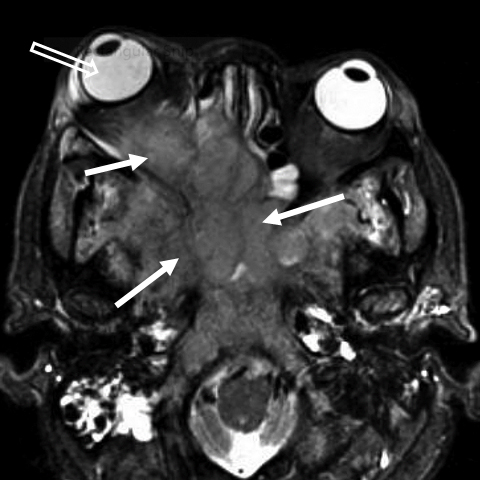
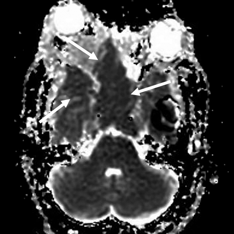
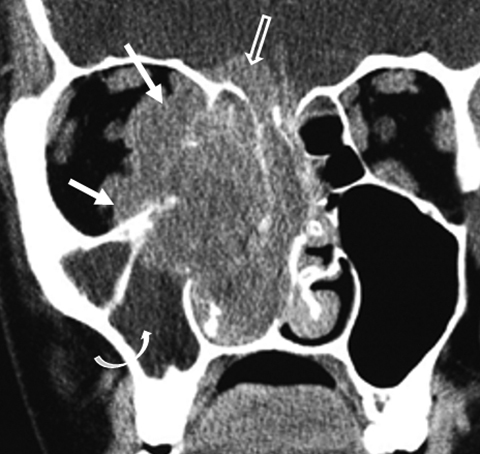
Non-Hodgkin lymphoma (NHL) is the most common non-epithelial neoplasm of the SN cavities and the B-cell type is most common in western countries. B-cell NHL typically presents in the 6th decade with no gender predilection.33 Lymphomas may be well-marginated or ill-defined and cause either osseous remodeling or destruction. Increased attenuation on CT, low signal intensity on long TR MR sequences, and diffusion restriction due to dense cellularity and increased nuclear to cytoplasmic (N:C) ratio within these tumors (Figure 20).32, 34 Variable, homogeneous enhancement is typical because of the homogeneous cell population.
Esthesioneuroblastoma (ENB)
Esthesioneuroblastoma is a neuroendocrine tumor that arises from the olfactory epithelium, and typically superiorly near the cribriform plate. The tumor has incidence peaks in adolescence and the 6th decade.35 Epistaxis is common due to the highly vascularity of this tumor. Long-term prognosis for ENB is excellent, with a 5-year survival of 75% despite orbital and intracranial extension.35 Imaging shows an avidly enhancing mass near the cribriform plate with destruction of the anterior skull base and/or lamina papyracea (Figure 21). With intracranial extension, the tumor may have a dumbbell shape with a “waist” at the level of the skull base. A distinguishing feature of ENB with intracranial extension is the formation of cysts at the tumor-brain interface.29, 36
Conclusion
Given the proximity of the SN cavities and orbits, it is not uncommon for patients with primarily SN pathology to present with ophthalmologic symptoms. Although some entities may be diagnosed clinically, recognizing the characteristic imaging features of these lesions when present is important. Although an exact histologic diagnosis may not be made, the pathology can almost always be classified as infectious/inflammatory, fibro-osseous or neoplastic. Granulomatous disease should be suspected in a patient with synchronous involvement of the SN cavities and the orbits. In addition to creating a reasonable differential diagnosis, identifying complications that may warrant a change in therapy is important for imagers.
References
- Sharma PK, Saikia B, Sharma R. Orbitocranial complications of acute sinusitis in children. J Emerg Med. 2014; 47(3):282-285.
- Rahbar R, Robson CD, Petersen RA, et al. Management of orbital subperiosteal abscess in children. Arch Otolaryngol Head Neck Surg. 2001; 127(3):281-286.
- Curtin HD, Rabinov JD. Extension to the orbit from paraorbital disease. The sinuses. Radiol Clin North Am. 1998; 36(6):1201-1213.
- Aribandi M, McCoy VA, Bazan C, et al. Imaging features of invasive and noninvasive fungal sinusitis: a review. Radiographics. 2007; 27(5):1283-1296.
- Glass D, Amedee RG. Allergic fungal rhinosinusitis: a review. The Ochsner Journal. 2011; 11(3):271–275.
- Marple BF. Allergic fungal rhinosinusitis: current theories and management strategies. Laryngoscope. 2001; 111(6):1006-1019.
- Loo JL, Looi AL, Seah LL. Visual outcomes in patients with paranasal mucoceles. Ophthal Plast Reconstr Surg. 2009; 25(2):126-129.
- Hourany R, Aygun N, Della Santina CC, et al. Silent sinus syndrome: an acquired condition. AJNR Am J Neuroradiol. 2005; 26(9):2390-2392.
- Tiegs-Heiden CA, Eckel LJ, Hunt CH, et al. Immunoglobulin G4-related disease of the orbit: imaging features in 27 patients. AJNR Am J Neuroradiol. 2014; 35(7):1393-1397.
- Fujita A, Sakai O, Chapman MN, et al. IgG4-related disease of the head and neck: CT and MR imaging manifestations. Radiographics. 2012; 32(7):1945–1958.
- Nwawka OK, Nadgir R, Fujita A, et al. Granulomatous disease in the head and neck: developing a differential diagnosis. Radiographics. 2014; 34(5):1240-1256.
- Morales-Angulo C, Garcia-Zornoza R, Obeso-Aguera S, et al. Ear, nose and throat manifestations of Wegener’s granulomatosis (granulomatosis with polyangiitis). Acta Otorrino laringol Esp. 2012; 63(3):206-211.
- Cannady SB, Batra PS, Koening C, et al. Sinonasal Wegener granulomatosis: A single-institution experience with 120 cases. Laryngoscope. 2009; 119(4):757–761.
- Demirci H, Christianson MD. Orbital and adnexal involvement in sarcoidosis: analysis of clinical features and systemic disease in 30 cases. Am J Ophthalmol. 2011; 151(6):1074–1080.
- Fergie N, Jones NS, Havlat MF. The nasal manifestations of sarcoidosis: a review and report of eight cases. J Laryngol Otol. 1999; 113(10):893-898.
- Erbek S, Erbek SS, Tosun E, et al. A rare case of sarcoidosis involving the middle turbinates: an incidental diagnosis. Diagn Pathol. 2006; 21;1:44.
- Brannon RB, Fowler CB. Benign fibro-osseous lesions: a review of current concepts. Adv Anatom Pathol. 2001; 8(3):126–143.
- Boudewyns AN, van Dinther JJS, Colpaert CG. Sinonasal fibro-osseous hamartoma: case presentation and differential diagnosis with other fibro-osseous lesions involving the paranasal sinuses. Eur Arch Otorhinolaryngol. 2006; 263(3): 276–281.
- McCarthy EF. Fibro-osseous lesions of the maxillofacial bones. Head Neck Pathol. 2013; 7(1):5-10.
- Sen S, Chandra A, Mukhopadhyay S, et al. Sinonasal tumors: computed tomography and MR imaging features. Neuroimaging Clin N Am. 2015; 25(4):595-618.
- Eversole R, Su L, ElMofty S. Benign fibro-osseous lesions of the craniofacial complex. A review. Head Neck Pathol. 2008; 2(3):177-202.
- Harrison DF. Osseous and fibro-osseous conditions affecting the craniofacial bones. Ann Otol Rhinol Laryngol. 1984; 93(3 Pt 1):199–203.
- Barnes L, Tse LLY, Hunt JL, et al. In: Barnes L, Eveson JW, Reichart P, Sidransky D, eds. Pathology and Genetics of Head and Neck tumors. Lyon, FR: IARC Press; 2005:9-80.
- Imola MJ, Schramm VL Jr. Orbital preservation in surgical management of sinonasal malignancy. Laryngoscope. 2002; 112(8 Pt 1):1 357-1365.
- Iannetti G, Valentini V, Rinna C, et al. Ethmoido-orbital tumors: our experience. J Craniofac Surg. 2005; 16(6):1085-1091.
- Llorente JL, López F, Suárez C, et al. Sinonasal carcinoma: clinical, pathological, genetic and therapeutic advances. Nat Rev Clin Oncol. 2014;11(8):460-472.
- Haerle SK, Gullane PJ, Witterick IJ, et al. Sinonasal carcinomas: epidemiology, pathology, and management. Neurosurg Clin N Am. 2013;24(1):39-49.
- Pilch BZ, Bouquot J, Thompson LDR. In: Barnes L, Eveson JW, Reichart P, Sidransky D, eds. Pathology and Genetics of Head and Neck Tumors. Lyon, FR: IARC Press; 2005:9-80.
- Das S, Kirsch CF. Imaging of lumps and bumps in the nose: a review of sinonasal tumours. Cancer Imaging. 2005; 9(5):167-177.
- Frierson HF Jr, Mills SE, Fechner RE, et al. Sinonasal undifferentiated carcinoma: an aggressive neoplasm derived from Schneiderian epithelium and distinct from olfactory neuroblastoma. Am J Surg Pathol. 1986; 10 (11):771–779.
- Wenig BM. Undifferentiated malignant neoplasms of the sinonasal tract. Arch Pathol Lab Med. 2009; 133(5):699-712.
- Eggesbø HB. Imaging of sinonasal tumours. Cancer Imaging. 2012; 12:136-152.
- Shohat I, Berkowicz M, Dori S, et al. Primary non-Hodgkin’s lymphoma of the sinonasal tract. Oral Surg Oral Med Oral Pathol Oral Radiol Endod. 2004; 97(3):328-331.
- Neves MC, Lessa MM, Voegels RL, et al. Primary non-Hodgkin’s lymphoma of the frontal sinus: case report and review of the literature. Ear Nose Throat J. 2005; 84(1):47-51.
- Bak M, Wein RO. Esthesioneuroblastoma: a contemporary review of diagnosis and management. Hematol Oncol Clin North Am. 2012; 26(6):1185-1207.
- Ow TJ, Bell D, Kupferman ME, et al. Esthesioneuroblastoma. Neurosurg Clin N Am. 2013; 24(1):51-65.
Citation
M A, MA M.Sino-orbital pathologies: An approach to diagnosis and identifying complications. Appl Radiol. 2017; (8):8-20.
August 1, 2017