Rupture of the Corona Mortis
Images
CASE SUMMARY
A 41-year-old male status post-motor vehicle accident presented with an open-book pelvic fracture. He was hemodynamically unstable. Imaging and subsequent treatment were performed in a hybrid Angio-CT suite (Canon Medical Systems USA, Inc.).
IMAGING FINDINGS
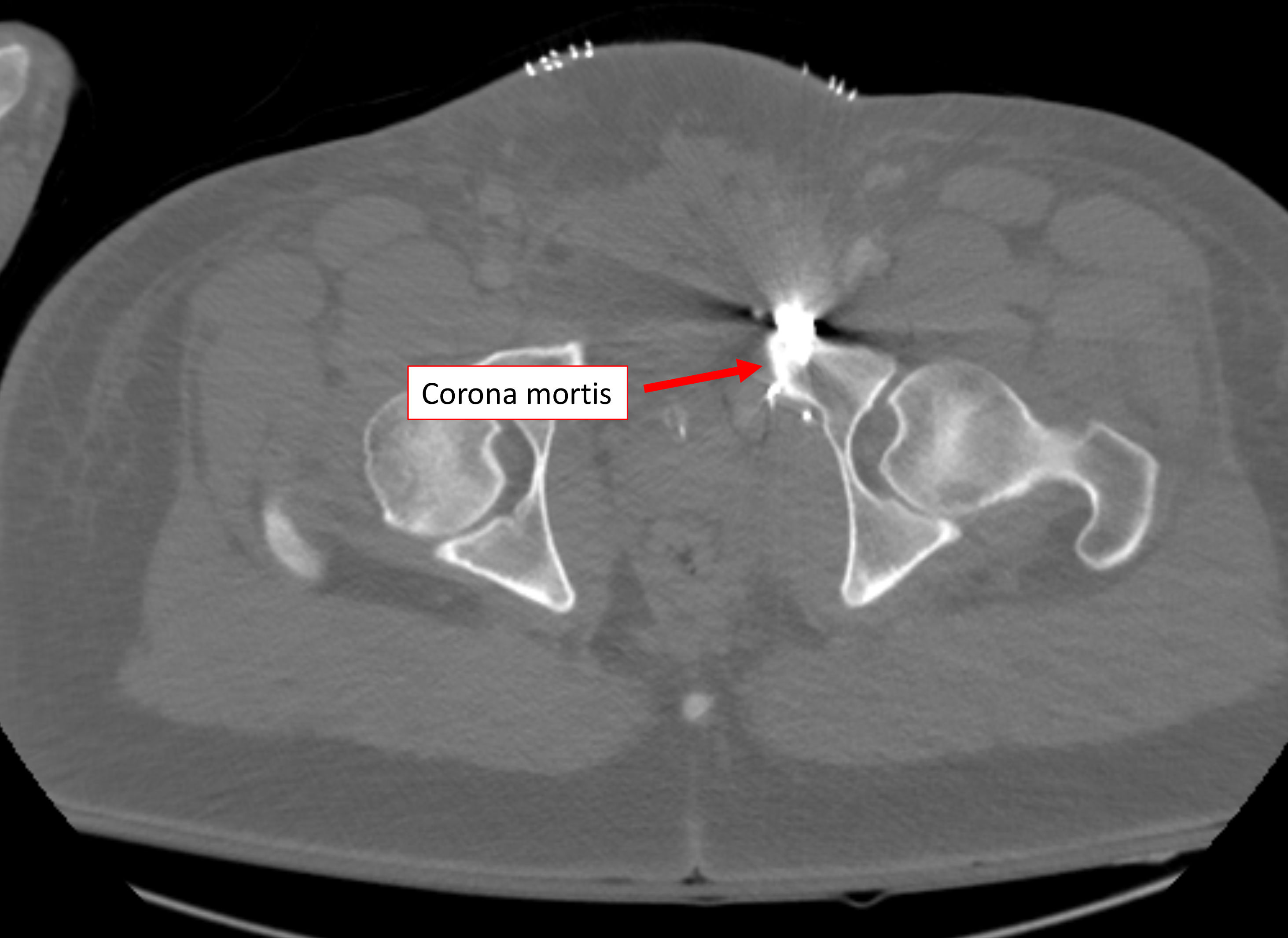
CT angiography of the pelvis revealed diastasis of the symphysis pubis and the left sacroiliac joint. A high-attenuation fluid collection, measuring 9.2 × 2.2 cm in the axial dimension and consistent for hematoma, was present in the subcutaneous fat, superficial to the lower-left rectus abdominis musculature.
Additional intermediate-to high-density fluid surrounded the rectus abdominis, displacing the bladder posterolateral to the right and extending craniocaudally. This fluid also had characteristics consistent with hematoma. No active extravasation of contrast was seen. Owing to continued hypotension, conventional angiography was performed.
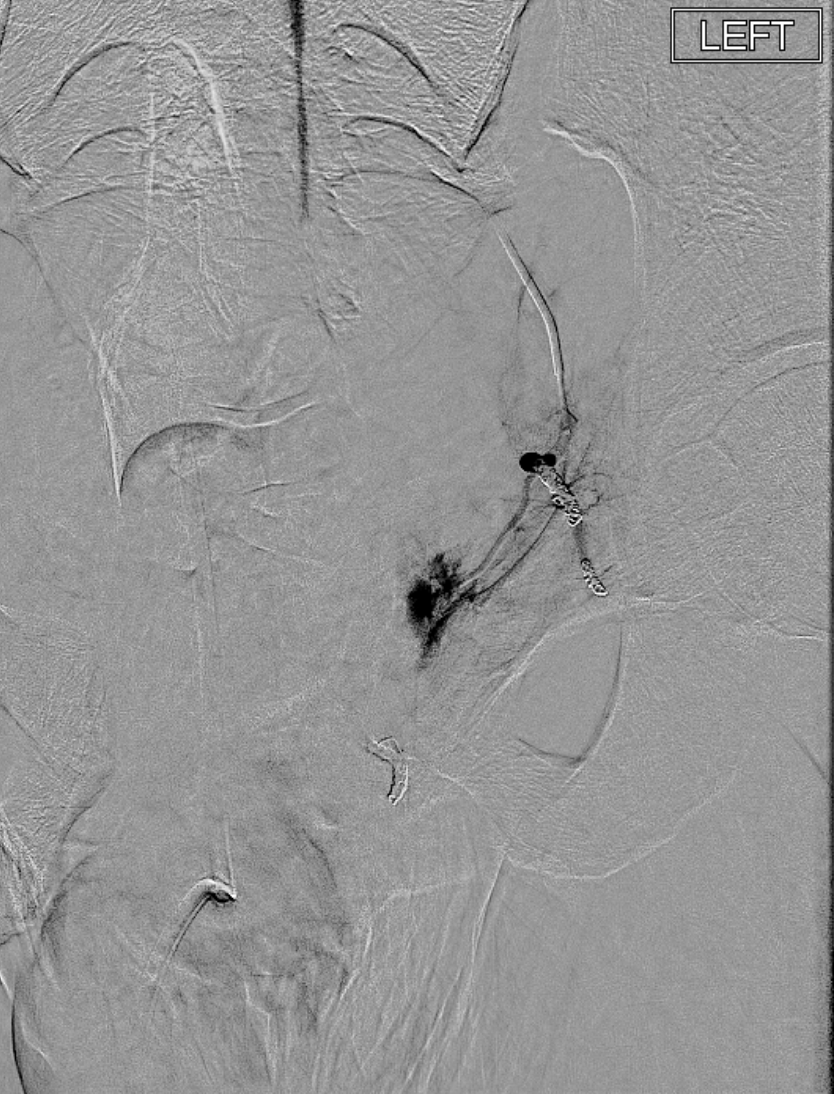
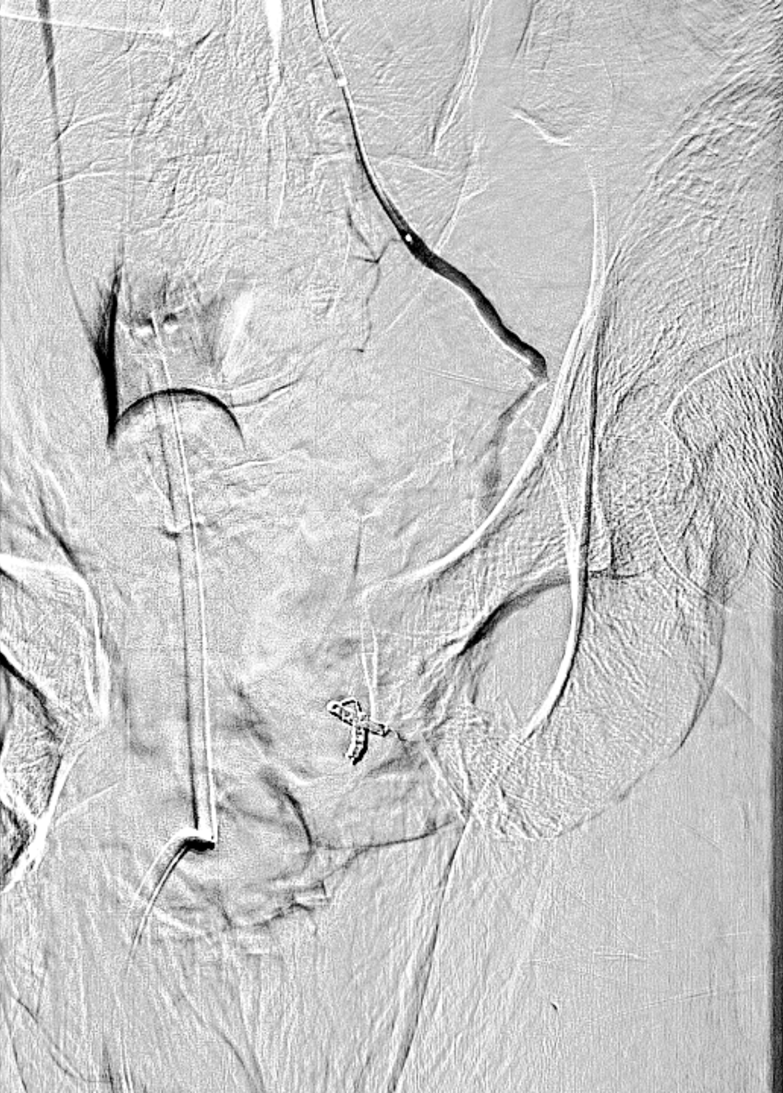
An initial non-selective pelvic angiogram demonstrated no active contrast extravasation to suggest active hemorrhage. There was marked lateral displacement of the left internal iliac artery and its branches.
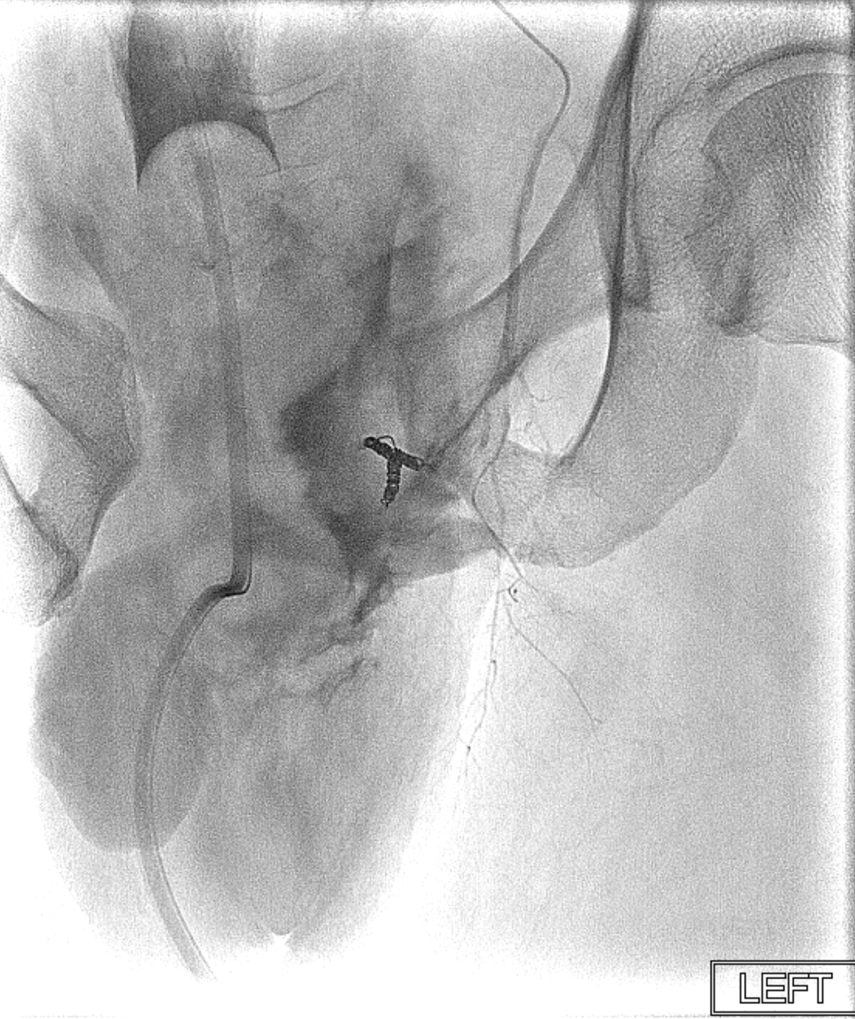
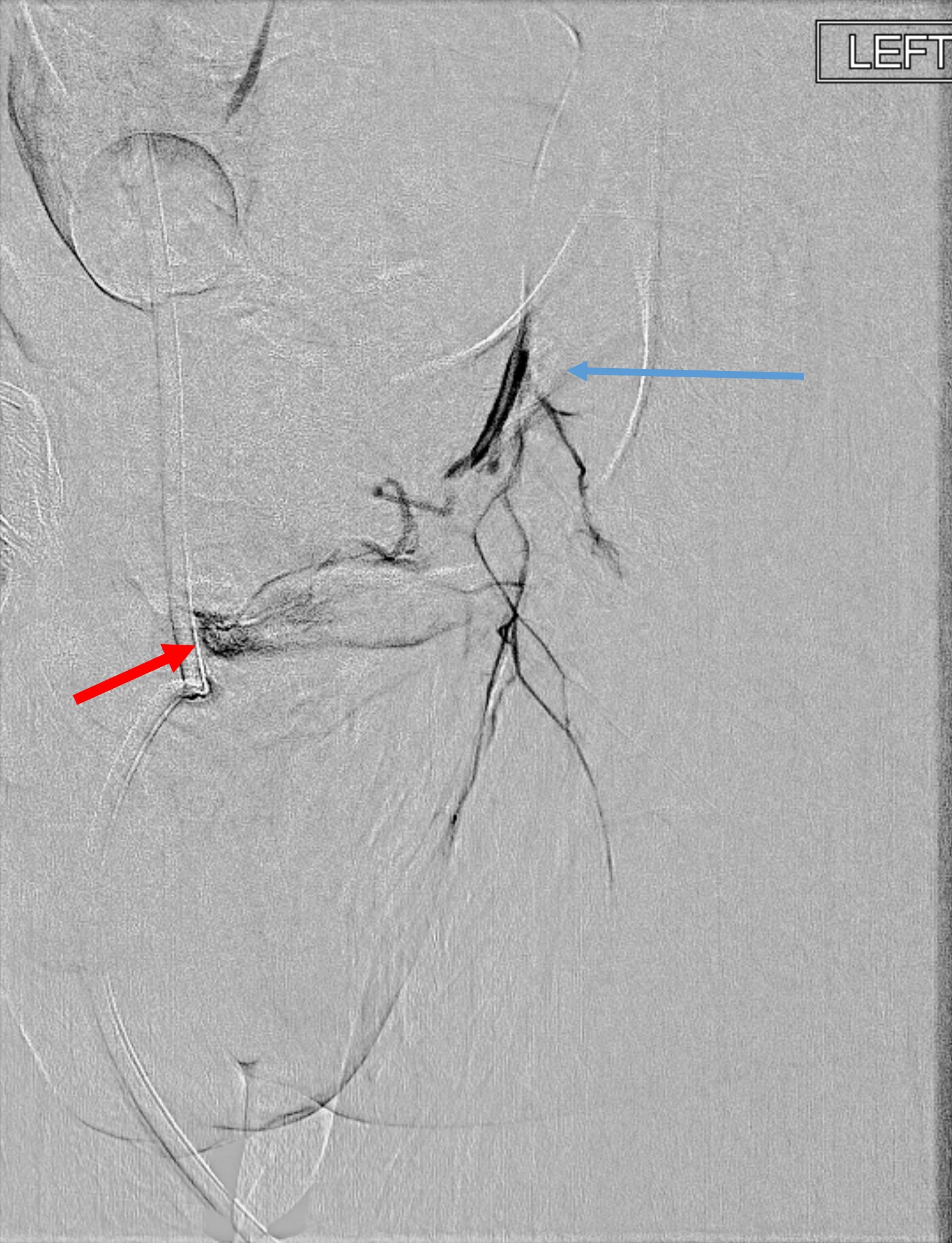
Super-selective catheterization of the left obturator artery demonstrated active contrast extravasation, which was embolized using gel foam and coils.
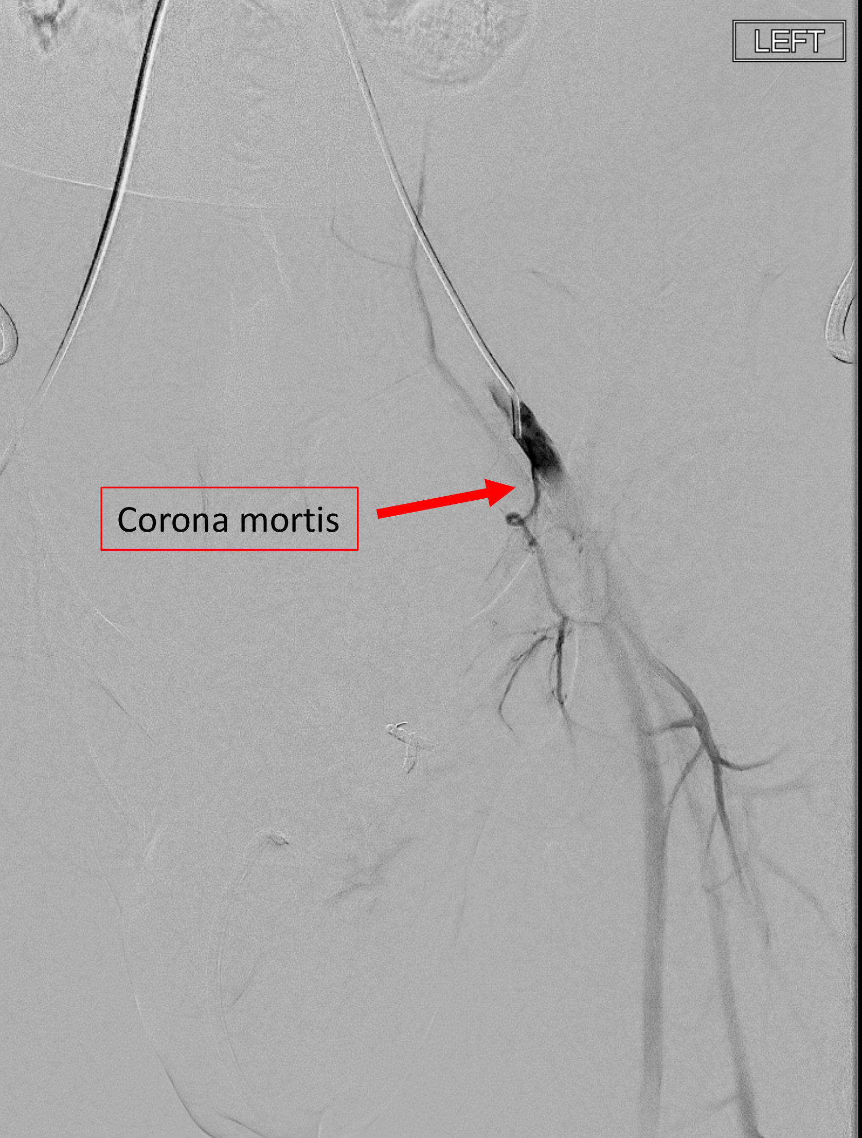
Selective angiogram of the left external iliac artery (EIA) revealed a corona mortis arising directly from the EIA. Super-selective catheterization revealed active extravasation. The corona mortis was embolized using micro-coils.
DIAGNOSIS
Rupture of the corona mortis
DISCUSSION
The corona mortis (“Crown of Death”) is a relatively common variant of the pelvic arterial vasculature that can be seen in up to one-third of patients. While numerous variations have been described, this variant most commonly exists as an artery arising from the external iliac or inferior epigastric artery that courses superficially to the pubis and joins with the obturator artery to form a ring or crown. Regardless of the specific origin of the communicating vessels, the underlying anatomy of an arterial segment coursing posterior to the superior pubic ramus makes it highly susceptible to injury in pelvic trauma and difficulty achieving hemostasis. Johnson et al identified external iliac branch injury in 17 of 66 patients (17%) with pelvic trauma, highlighting the importance of interrogating this vascular bed.
While most radiologists and trauma surgeons can recall the corona mortis when prompted, it can easily be forgotten while rendering emergent care to an unstable trauma patient. Overlooking a corona mortis, in keeping with its name, may be the difference between life and death.
Corona mortis injuries can easily be missed unless systematically excluded. Therefore, conventional practice when assessing a hemodynamically unstable patient with pelvic trauma, especially an open-book fracture, is to evaluate the internal and external iliac arteries with pelvic angiography.
Access to a hybrid Angio-CT suite, where diagnostic CTA followed by conventional angiography and embolization can be performed on the same table eliminates patient transfer and reduces the time for successful treatment versus traditional approaches. Further, the hybrid Angio-CT suite may also expedite this process, as diagnostic CTA can definitively identify the corona mortis and permit detailed planning prior to embolization.
CONCLUSION
The corona mortis is a relatively common vascular variant that can be overlooked in the setting of pelvic trauma. Prompt identification and treatment are necessary to avoid associated morbidity and mortality. Angio-CT may represent a viable new method to streamline recognition of and intervention for this significant finding.
References
- Berberoğlu M, Uz A, Ozmen MM, et al. Corona mortis: an anatomic study in seven cadavers and an endoscopic study in 28 patients. Surg Endosc. Jan 2001;15(1):72-5. doi:10.1007/s004640000194
- Darmanis S, Lewis A, Mansoor A, Bircher M. Corona mortis: an anatomical study with clinical implications in approaches to the pelvis and acetabulum. Clin Anat. May 2007;20(4):433-9. doi:10.1002/ca.20390
- Johnson GE, Sandstrom CK, Kogut MJ, et al. Frequency of external iliac artery branch injury in blunt trauma: improved detection with selective external iliac angiography. J Vasc Interv Radiol. Mar 2013;24(3):363-9. doi:10.1016/j.jvir.2012.12.006
- Perandini S, Perandini A, Puntel G, Puppini G, Montemezzi S. variant of the obturator artery: a systematic study of 300 hemipelvises by means of computed tomography angiography. Pol J Radiol. 2018;83:e519-e523. doi:10.5114/pjr.2018.81441
- Pua U, Teo LT. Prospective diagnosis of corona mortis hemorrhage in pelvic trauma. J Vasc Interv Radiol. Apr 2012;23(4):571-3. doi:10.1016/j.jvir.2011.12.018
- Smith JC, Gregorius JC, Breazeale BH, Watkins GE. The corona mortis, a frequent vascular variant susceptible to blunt pelvic trauma: identification at routine multidetector CT. J Vasc Interv Radiol. Apr 2009;20(4):455-60. doi:10.1016/j.jvir.2009.01.007
- Steinberg EL, Ben-Tov T, Aviram G, Steinberg Y, Rath E, Rosen G. Corona mortis anastomosis: a three-dimensional computerized tomographic angiographic study. Emerg Radiol. Oct 2017;24(5):519-523. doi:10.1007/s10140-017-1502-x
Citation
. Rupture of the Corona Mortis . Appl Radiol.
March 19, 2021