Researchers Visualize Blood Flow Making Waves Across the Surface of a Mouse Brain
Images

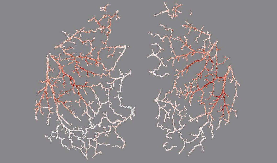
For the first time, researchers have visualized the full network of blood vessels across the cortex of awake mice, finding that blood vessels rhythmically expand and contract leading to “waves” washing across the surface of the brain. These findings, funded by the National Institutes of Health (NIH), improve the understanding of how the brain receives blood, though the function of the waves remains a mystery.
A network of elastic and actively pumping vessels carrying oxygenated blood span the surface of the brain before entering the cortex. There, they feed into a second network of capillaries that supply oxygen deeper into the tissue. Using physics-based experimental methods and analyses, the researchers saw that in addition to the pulses of blood flow that occur with each heartbeat, there are slower waves of blood flow changes that sweep across the brain and occur about once every ten seconds. The change in blood flow that occur with these slow waves was up to 20% of the entire brain blood supply. Surprisingly, this phenomenon was only weakly tied to changes in brain activity.
The waves produced visible bulges in the blood vessels, which will aid in mixing the fluid around the brain’s cells. This has implications in how waste products and other materials are removed from the fluid surrounding brain cells. Because the waves of bulging blood vessels move in a variety of directions, the authors surmise that the pulses of dilation and contraction of the blood vessels are more likely to be involved in mixing the fluid around them rather than actively moving it in a given direction. Regardless, this mixing activity could aid in removing misfolded proteins and other components from the brain into the cerebrospinal fluid that surrounds it. This process is considered an important protective mechanism for a variety of neurological disorders, such as Alzheimer’s disease and other related dementias, and is more active during sleep.
These findings may also affect current approaches to interpreting fMRI scans, which measure changes in blood oxygenation within brain structures as they are activated. Specifically, the finding that these waves of blood flow changes occur largely independent of brain activity suggests a new level of complexity complication that needs to be considered when interpreting the link between the fMRI data and brain activation.