Radiological Case: Multifocal osteomyelitis
Images


CASE SUMMARY
A 57-year-old confused Hispanic man presented to the emergency department with a 3-day history of leg pain. His past medical history was significant for epilepsy, alcoholism and psychosis. The physical exam revealed limited range of motion and pain of the left hip and knee. Admitting labs demonstrated a mild leukocytosis. Contrast-enhanced CT (CECT) scans of the pelvis and chest were obtained during the hospital stay.
IMAGING FINDINGS
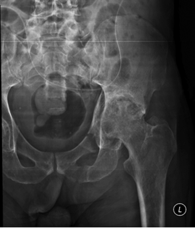
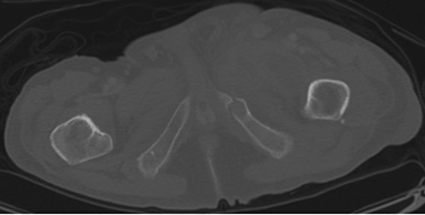
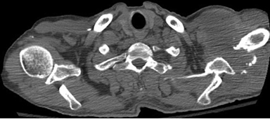
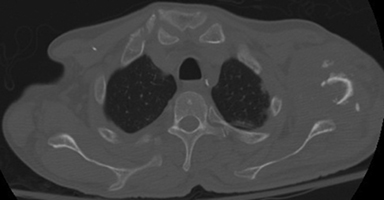
An initial radiograph preformed in the emergency department revealed a left pubic rami fracture (Figure 1). A follow-up CECT of the pelvis demonstrated a left superior pubic rami fracture of indeterminate age and a left inferior pubic rami fracture with a lytic appearance and expansion of the bone. In addition, the CT showed a large multiseptated and multiloculated, hypodense fluid collection with a few tiny air bubbles seen in the nondependent portion of the collection. The collection coursed between the fractures toward the left inguinal area within the adductor muscle group (Figure 2). On the second hospital day, the patient became diaphoretic and tachypenic, and a CT angiography of the chest was performed to evaluate for a pulmonary embolism. The CT showed a left humeral lytic expansile and destructive bone lesion with an associated soft-tissue component extending from the axilla to the left supraclavicular region (Figure 3).
DIAGNOSIS
Multifocal osteomyelitis with associated soft-tissue abscesses. Culture positive for Staphylococcus aureus.
Differential diagnosis: Bone metastasis.
DISCUSSION
Osteomyelitis is an inflammation of the bone typically caused by infection. Staphylococcus is responsible for most cases of osteomyelitis. 1 Chronic osteomyelitis results from a longstanding infection, on average longer than 6 weeks.2 Chronic osteomyelitis typically results in sclerosis. 3 Our patient, however, had lytic destruction, not sclerotic destruction. The differential diagnosis for multiple lytic lesions in an adult is metastatic disease or multiple myeloma.4 An infectious process such as osteomyelitis is not usually included in the differential diagnosis. In addition, it is uncommon for an adult to have multiple areas affected by ostemyelitis, as seen in our patient, compared to the pediatric population.5
Our patient had no history of immune deficiency disorders, sickle cell anemia, IV drug abuse, diabetes mellitus or subacute bacterial endocarditis, which all increase the risk of multifocal bacterial osteomyelitis.3 Pathological fractures are not common in osteomyelitis, especially in cases without devastating septicemia. 6 Our patient had negative blood cultures throughout his 10-day hospital stay. Our patient also had no prior history of osteomyelitis, which has been theorized to increase the risk of pathological fractures.6 The source of our patient’s initial infection is unknown. It is uncertain if our patient had osteomyelitis leading to the multiple soft-tissue abscesses, or if the soft-tissue abscesses led to the osteomyelitis. It is also unclear why the bony infections led to lytic destruction of the humeral head and pathological expansile fractures of the pubic rami in such a short time (less than 2 years).
CONCLUSION
Our patient had an uncommon presentation of multifocal osteomyelitis. Multifocal osteomyelitis typically occurs in the pediatric population, and the affected bone does not usually mimic metastatic lytic metastases. CECT imaging is useful in evaluating lytic bone lesions with associated soft-tissue abcesses. Histopathological correlation is key to determining malignancy or atypical cells. It is important to consider osteomyelitis in the differential diagnosis despite unusual presentation to ensure prompt administration of intravenous antibiotics.
REFERENCES
- Stalcup ST, Pathria MN, Hughes, TH. Musculoskleletal infections of the extremities: A tour from superficial to deep. Applied Radiology. 2011;40(11) retrieved from http://www.appliedradiology.com/articles/musculoskeletal-infections-of-the-extremities-a-tour-from-superficial-to-deep.
- Boutin RD, Brossmann J, Sartoris DJ, et al. Update on imaging of orthopedic infections. Orthopedic Clinics of North America.1998;29(1):41-66.
- Gold RH, Hawkins RA, Katz RD. Bacterial osteomyelitis: Findings on plain radiography, CT, MR and Scintigraphy. Am J Radiol. 1991;157: 365-370.
- Bennett DL, El-Khoury GY. General approach to lytic bone lesions. Applied Radiology. 2004;33(5): 8-17.
- Kothari NA, Pelchovitz DJ, Meyer JS. Imaging of musculoskeletal infections. Radiologic Clinic of North America. 2001;39(4): 653-671.
- Walmsley BH. Pathological fracture in acute osteomyelitis in an adult. J Royal Soc Med. 1982;76:77-78.
Citation
Radiological Case: Multifocal osteomyelitis. Appl Radiol.
September 2, 2015