Radiological Case: Diabetic myonecrosis
Images


CASE SUMMARY
A 51-year-old woman presented to the emergency room with spontaneous severe left thigh pain and mild left thigh swelling. Significant medical history included uncontrolled diabetes mellitus, coronary artery disease with stent placement, hyperlipidemia, and hypertension.The patient denied recent trauma or intramuscular injections. The patient’s laboratory findings included a normal CBC; elevated glucose of454 mg/dl; elevated Hgb A1c of 16.4%; and a normal CK level of 70 u/l. A venous ultrasound taken upon admission was negative for deep vein thrombosis (DVT). The patient was placed on antibiotic therapy for suspected cellulitis with no clinical improvement. A magnetic resonance imaging (MRI) scan was subsequently obtained.
IMAGING FINDINGS
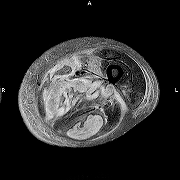
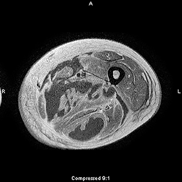
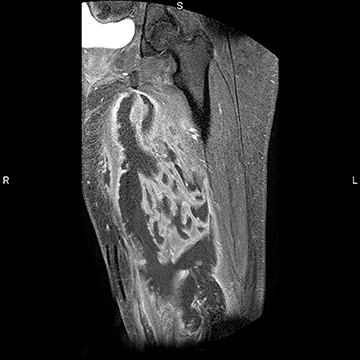
As a ligament injury was suspected, T2-weighted images (Figure 1) showed extensive intramuscular fluid within the adductor and hamstring muscle groups, and intramuscular edema within the quadriceps muscle. Diffuse fascial edema and subcutaneous soft-tissue edema was also noted. T1-weighted, postcontrast images (Figures 2 and 3) showed intense muscular rim enhancement outlining areas of muscle necrosis within the adductor and hamstring muscle groups.
DIAGNOSIS
Posterior ankle impingement syndrome due to os trigonum syndrome
DISCUSSION
Diabetic myonecrosis, or diabetic muscle infarct (DMI), is an uncommon and underdiagnosed complication of diabetes mellitus(DM). DMI is generally self-limiting, requiring only conservative treatment. However, failure to recognize this condition can result insignificant morbidity. DMI is often misdiagnosed as abscess, myositis, necrotizing fasciitis, or neoplasm.1 DMI should be considered in the differential of acute muscle pain in patients with DM.
First described in 1965, DMI is often seen in longstanding diabetics. A significant proportion of patients are noncompliant with their insulin regimens and have poor diabetic control. DMI is unusual as it occurs more frequently in young adults, with a female predilection.Patients present with acute onset of severe pain and swelling in the absence of fever or trauma. Other signs of inflammation are usually absent. The quadriceps compartment is most frequently involved, followed by the thigh adductors and hamstrings. Calf involvement has also been reported. Bilateral involvement may be seen in 8% to 10% of cases.2 Patients are known to have recurrences, usually within 2months of initial presentation. Laboratory tests typically show normal white cell count and normal or slightly elevated levels of creatine kinase. The condition’s pathogenesis is unknown, but is assumed to be from microvascular disease.
Clinically, DMI must be differentiated from other conditions that cause acute leg pain in diabetic patients, including DVT, superficial thrombophlebitis, cellulitis, necrotizing fasciitis, soft-tissue abscess, soft-tissue hematoma, and tumor.
MRI plays an important role in diagnosing DMI. The main findings are increased intramuscular signal on T2-weighted sequences.Affected muscles are either hypo- or isointense on T1-weighted sequences. Foci of high signal on T1 images may indicate intramuscular hemorrhage. Affected muscles show modest gadolinium enhancement. Muscle infarction is depicted by muscle-rim enhancement.Other findings include diffuse enlargement of affected muscles and indistinct muscle planes. Subcutaneous edema and parafascial fluid are also commonly seen on T2-weighted sequences. Sonographic findings of DMI include internal linear echogenic structures coursing throughout the muscles, without a sonographically detectable abscess or mass. Radiological differential diagnosis of DMI includes soft tissue abscess, pyomyositis and necrotizing fasciitis. Pyomyositis and necrotizing fasciitis may not be distinguishable from DMI solely on the basis of MRI findings. Patients with pyomyositis and necrotizing fasciitis are more likely to have fever, cellulitis and an elevatedWBC count. Severe pain is very characteristic of DMI and may not be seen with necrotizing fasciitis or pyomyositis.3
CONCLUSION
DMI is a rare, underdiagnosed complication of DMI. Patients with DMI often present with severe thigh pain or swelling. DMI should always be considered in the differential diagnosis of acute muscle pain in a diabetic patient. Early noninvasive diagnosis is possible with MRI. DMI often only requires conservative management and minimal intervention. Biopsy is rarely indicated. The diagnosis remains a marker of severe uncontrolled diabetes. Aggressive control of blood sugar is imperative, as many patients with DMI have an elevated5-year mortality due to other diabetic complications.4
REFERENCES
- Wintz RL, Prinstone KR, Nelson SD. Detection of diabetic myonecrosis: Complication is often missed sign of underlying disease. Postgrad Med. 2006;119:66-69.
- Trujillo-Santos AJ: Diabetic muscle infarction: An underdiagnosed complication of longstanding diabetes. Diabetes Care. 2003;26:211-215.
- Delaney-Sathy LO,Fessell DP, Jacobson JA, Hayes CW. Sonography of diabetic muscle infarction with MR imaging and pathologic correlation. AJR. Am J Roentgen. 2000;174:165-169.
- Meher D, Mathew V, Misgar R, et al. Diabetic myonecrosis: An Indian experience. Clin Diabetes. 2013;31:53-58.
Citation
Radiological Case: Diabetic myonecrosis. Appl Radiol.
February 6, 2014