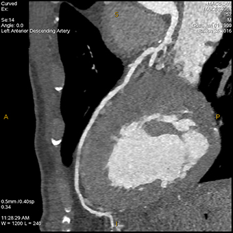
Practical Guide Published for CT Fractional Flow Reserve
 Radiographics has published a practical guide for the application and interpretation of CT fractional flow reserve (FFRCT) that address common pitfalls and challenges. The journal based SA-CME activity from the Mayo Clinic also provides techniques and practical tips for the imaging exam “that improves the specificity of coronary CT angiography (CTA) in the evaluation of CAD by providing the hemodynamic significance of a stenotic lesion,” the authors conclude.
“CT Fractional Flow Reserve: A Practical Guide to Application, Interpretation, and Problem Solving,” provides guidance on interpretation, where FFRCT should be measured, identifying at-risk patients, addressing discordant values and more. Lesion-specific ischemia using FFRCT should be measured 2 cm distal to the stenotic lesion. The authors provide values for evaluating the lesion, with 0.75 or less being abnormal, 0.75-0.8 being borderline and greater than 0.8 being normal. Clinical and anatomical coronary CTA findings should be correlated to FFRCT.
According to the article, cases with mild stenosis can result in abnormal FFRCT values, cases with severe stenosis can result in normal FFRCT values and gradually decreasing or abnormally low FFRCT values at the distal vessel without a proximal focal lesion could be due to diffuse atherosclerosis.
In addition to acting as a “gatekeeper” for invasive coronary angiography (ICA) by decreasing its use in patients with nonobstructive disease, FFRCT improves the specificity of coronary CTA by evaluating lesion-specific ischemia. The authors note the importance of interpreting FFRCT along with other factors such as symptoms, clinical history, comorbidities, location of the abnormal FFRCT, coronary anatomy and presence of revascularization targets.
Related Articles
Citation
. Practical Guide Published for CT Fractional Flow Reserve. Appl Radiol.
February 9, 2022