Peripheral vascular CTA: Emerging role of PV-CTA in the therapeutic management of PVD
Images
Dr. Allie is Chief of Cardiothoracic and Endovascular Surgery, Dr. Patlola is an Interventional Cardiologist, Dr. Ingraldi is an Interventional Cardiologist, Mr. Hebert is Director of Cardiovascular Services, and Dr. Walker is the Medical Director, Founder and President, Cardiovascular Institute of the South, Lafayette, LA. Mr. Hebert is also the Director of the Cardiac Cath Lab, Southwest Medical Center, Lafayette, LA.
Disclosures: Dr. Allie and Dr. Walker serve as Consultants to Toshiba, Bracco Diagnostics, and The Spectranetics Corporation. Dr. Patlola and Dr. Ingraldi have nothing to disclose. Mr. Hebert is a Consultant to The Spectranetics Corporation.
It is estimated that there are 15 to 20 million patients with peripheral vascular disease (PVD) in the United States. 1,2 Likewise, there are 20 million patients with diabetes mellitus and a similar number of patients with peripheral venous disease. These patient populations make the clinical opportunity in peripheral vascular (PV) computed tomographic angiography (CTA), or PV-CTA, even greater than it is for cardiac CTA. 1,2 It is estimated that 3 to 4 million symptomatic PVD patients are misdiagnosed or go untreated and that twice that number are asymptomatic and therefore untreated. 1,3 This asymptomatic group includes patients with abdominal aortic aneurysm (AAA), significant internal carotid artery (ICA) and vertebral artery disease, renal artery stenosis (RAS), and mesenteric vascular disease. Peripheral vascular CTA is the ideal noninvasive tool to identify this large, asymptomatic, yet at-risk patient population for whom endovascular therapeutic treatments are now available. Therefore, earlier PVD diagnosis and treatment facilitated by PV-CTA have the potential to significantly improve patient outcomes.
Conventional angiography with digital subtraction angiography (DSA) remains the gold standard for vascular imaging but has multiple limitations. Magnetic resonance angiography (MRA) has been advocated to address the limitations of DSA, but MRA also possesses significant limitations. This article reviews the contemporary utilization of PV-CTA and describes the authors' experience with 64-channel PV-CTA. CTA has the potential to become a paradigm shift in not only the diagnostic, but also the overall therapeutic management of PVD.
PV-CTA imaging technique
Current 64-channel CTA imaging acquisition and injection parameters are primarily tailored to cardiac and coronary pathology. In clinical CTA, the contrast enhancement, acquisition parameters, coverage speed, and circulating time are accurately determined by protocols that are based on the clinical information requested, the imaging modality used, and the patient-specific variables (age, body size, cardiac output, renal function, etc.). Therefore, these rapid scan and acquisition times oftentimes are not optimal for PVD. Optimal contrast enhancement and image acquisition in PVD patients require 64-channel CTA injection parameter adjustments to "slow down" or "time" the data acquisition to the slower blood flow rates in the PVD patient, especially those with significant infrapopliteal disease. Our group has reported low-contrast 64-channel CTA protocols in patients with PVD and critical limb ischemia (CLI). 4,5
The PV-CTA protocol also serves as our CLI-CTA protocol by adding a second below-the-knee scan for infrapopliteal imaging enhancement. We reported our results of a comparison of DSA versus CTA in 140 PVD and 98 CLI patients. The DSA versus CTA % stenosis correlations were strong and included: carotid (r 2 = 0.957), renal (r 2 = 0.942), celiac (r 2 = 0.907), mesenteric (r 2 = 0.909), iliac (r 2 = 0.951), superior femoral (r 2 = 0.903), popliteal (r2 = 0.889), peroneal (r 2 = 0.882), anterior tibial (r 2 = 0.907), and posterior tibial (r 2 = 0.891). 4,5 Integral to these PV-CTA protocols are the higher iodinated contrast agents, injection parameter changes, second lower extremity scans, and low contrast volume while facilitating postprocessing resolution and image enhancement.
Clinical PV-CTA applications
Carotid artery disease
Conventional angiography with DSA remains the gold standard for ICA imaging but has a definite risk of periprocedural stroke. Digital subtraction angiography is performed in limited projections, while CTA provides multiple3-dimensional (3D) views. Digital subtraction angiography has been shown to underestimate the degree of ICA stenosis when surgical specimens of cross-sectional lumens were compared with the results of DSA. 6 Using helical CTA, Elgersma et al 7 identified additional ICA (16%) suitable for carotid endarterectomy (CEA) compared with DSA, therefore further underscoring the complexity of the ICA bifurcation and the need for multiple views to accurately determine the degree of stenosis. Likewise, mounting evidence suggests that carotid plaque morphology characteristics are associated with outcomes and clinical events. 7,8 Carotid CTA holds promise in facilitating this determination and, therefore, has significant clinical therapeutic treatment implications.
Carotid duplex ultrasound (DU) has been known to have >90% sensitivity and specificity in diagnosing ICA disease but is still operator-dependent, moderately time-consuming, and gives information primarily only on ICA bifurcational disease. Several reports have shown the diagnostic accuracy of carotid CTA in cases of 70% to 99% ICA stenosis to have a sensitivity of 100% and specificity of 94% to 100%. 9- 11 Multiplanar reconstruction (MPR) methods will allow cross-sectional luminal and plaque morphology evaluation and will provide additional pertinent clinical information. 12
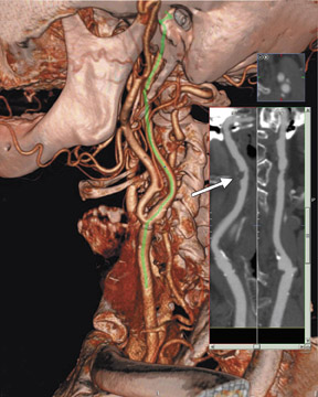
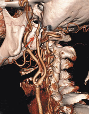
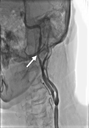
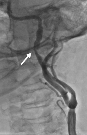
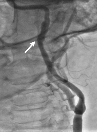
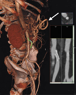
Aortic arch vessel anatomy, tortuosity, and ostial arch vessel disease along with distal ICA tortuosity are often significant limitations to carotid artery stenting (CAS), since placement of a distal protection device (DPD) is now considered the standard of care. 13 CTA allows accurate assessment of ICA stenosis, access vessel and distal ICA tortuosity, and the detection of arch vessel disease, thus potentially allowing a determination of CAS versus CEA candidacy noninvasively without the inherent risks of DSA. 13,14 Excellent proximal vertebral artery imaging is available on a routine carotid CTA, allowing identification of additional symptomatic patients with vertebral artery symptoms who could benefit from vertebral artery percutaneous transluminal angioplasty (PTA) or stenting. Carotid CTA has replaced traditional angiography as our carotid system diagnostic tool of choice, and increasingly important carotid therapeutic treatment and surveillance decisions are made relying solely on CTA information (Figure 1).
Renal artery disease
The benefits of RAS diagnosis and revascularization have been proven in patients with severe hypertension, refractory congestive heart failure, or angina, and in patients with declining renal function, which underscores the importance of a simple accurate, noninvasive diagnostic tool. The limitations of renal artery DU are well known. Three-dimensional abdominal CTA has addressed many of these limitations, and Beregi et al 15 recently reported 100% accuracy and sensitivity comparing the use of CTA with DSA in RAS. Galanski et al 16 reported the sensitivity and specificity of CTA versus DSA in RAS as 92% and 95%, respectively. Abdominal CTA is rapidly replacing DU and MRA as the primary diagnostic and surveillance tool in evaluating RAS, especially for in-stent restenosis (ISR). In patients with RAS, the CTA scanner can pinpoint the juxtarenal aorta and shorten acquisition times to <10 seconds and reduce contrast load to <40 mL.
The incidence of renal artery ISR has been reported to be as high as 15% to 20% and has been associated with vessel size and stent undersizing. 17 Accurate preprocedural knowledge of vessel anatomy and size may facilitate more precise PTA/stenting and thus decrease ISR. Plaque morphology and branching anatomy may also facilitate decisions regarding DPD use, especially in ulcerative lesions. Peripheral vascular CTA provides an excellent imaging tool for the diagnosis of and treatment planning for renal artery ISR.
Mesenteric artery disease
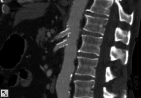
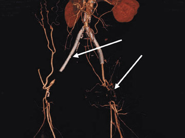
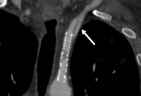
Asymptomatic and symptomatic mesenteric artery disease (MAD) (celiac and superior-inferior mesenteric artery disease) has been difficult to diagnose, both clinically and by traditional imaging techniques; therefore, MAD is underappreciated, underdiagnosed, and undertreated. This is likely analogous to our understanding and treatment of RAS 2 decades ago. Recent reports have identified the benefits of PTA/stenting in appropriately diagnosed patients with MAD. 18,19 There are no reports of 64-channel CTA evaluating MAD, but our initial experience indicates that CTA will be as accurate in diagnosing MAD as it is in RAS. Stents are well imaged, allowing the potential for improved accuracy in detecting MAD ISR, which, like RAS, has a 15% to 20% incidence 18,19 (Figure 2).
An abdominal aorta CTA with distal runoff begins acquiring images at the diaphragm level and thus will potentially identify a significant incidence of MAD. It is likely the incidence of MAD is as common as RAS and, therefore, will be a new vascular territory available for PTA/stenting. Traditionally, the treatment of MAD has required difficult major vascular surgical reconstructions with high mortalities and morbidities. The endovascular MAD treatment techniques are very similar to treating RAS. 18,19 It is likely that with CTA and endovascular treatments, the incidence of MAD treatment will increase in the near future with improved outcomes.
Aortoiliac occlusive disease
Several studies have shown the sensitivity and specificity of CTA in detecting significant aortoiliac occlusive disease (AIOD) to be >96%. 20 Rubin et al 21 recently reported 100% concordance between CTA and DSA in AIOD using 2.5-mm slices covering 120 mm in a 30-second acquisition time. Digital subtraction angiography can fail to detect aneurysmal disease, and the diagnostic accuracy is adversely affected by vascular calcification. Single planar DSA frequently "misses" eccentric lesions, which are prevalent posteriorly at the distal aortic bifurcation and the entire iliac artery segment. CTA has been shown to be superior to DSA in evaluating vascular trauma, dissections, and aneurysms. Recent reports have shown a reduction in contrast use and a 4-fold reduction in radiation exposure when comparing CTA with DSA for the diagnosis of AOID. 22 Several asymptomatic >5.0-cm AAAs and significant iliac aneurysms are diagnosed each month at our institute in patients who are imaged for suspected occlusive disease.
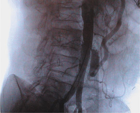
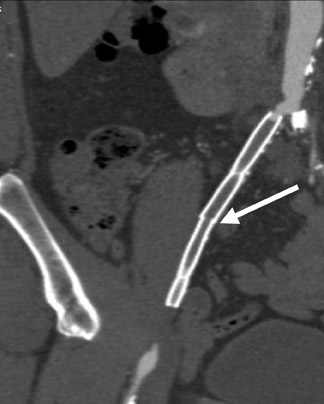
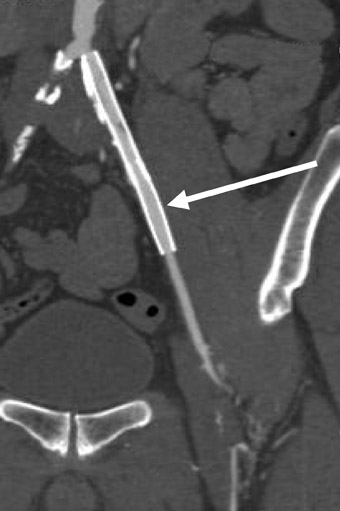
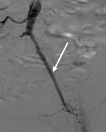
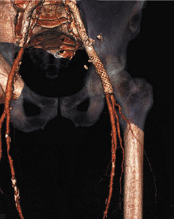
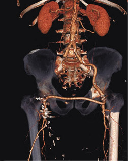
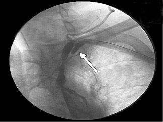
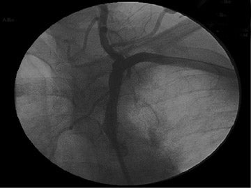
Our routine CTA for AIOD includes cephalocaudad coverage from the supra-celiac aorta to the proximal thigh (approximately 30 to 40 cm). Native common femoral artery (CFA) disease is not uncommon, and frequently patients will present with "stick site injuries" from multiple prior procedures. Avoiding a "diagnostic stick" with CTA versus diagnostic DSA has significant clinical implications. We have found CTA to be particularly useful in avoiding "sticking into CFA disease" or "stick site injuries" (Figure 3). It is always recommended to scan both legs, since most endovascular procedures are performed via a CFA approach.
Abdominal aortic aneurysm
Since the introduction of aortic stent grafts and endovascular aneurysm reconstruction (EVAR), CTA has rapidly become the gold standard for abdominal aortic pathology diagnosis, treatment planning, and postoperative surveillance. CTA offers several potential advantages over traditional imaging, including: 1) superiority in identifying mural thrombus and in evaluating periaortic tissues for rupture, endoleak, and inflammatory AAA identification; 2) more precise determinations of size, length, angulation, and transverse dimensions of the AAA superior neck; 3) more accurate characterization of postprocedural juxtarenal AAA endograft deformation, kinking, or migration with identification of branch vessels; 4) 3D reconstruction that allows improved assessment of iliac tortuosity and detection of endoleaks; and 5) post-EVAR AAA volume determinations that may allow earlier detection of device failure, rupture, or endoleak.
Infrainguinal disease
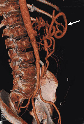
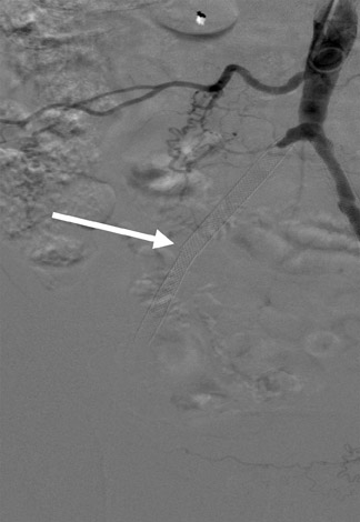
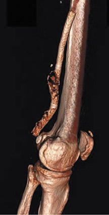
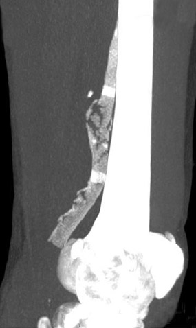
The femoral and infrapopliteal vessels are among the most diseased and calcified vessels in the body and are thus a challenge for vascular imaging. There are no published data that compare DSA or DU with 64-channel CTA in infrainguinal vessels, and few data that evaluate 4- to 16-chan-nel CTA. 23 Several promising image enhancement-processing techniques are available, especially for infrapopliteal vessels. Curved planar reformation (CPR) and semitransparent volume rendering (STVR) with automated measurements are new 3D imaging modalities that improve the accuracy of CTA in highly calcified vessels. The editing of boney structures (osseous segmentation) is now available at the workstation with the use of automatic region-growing imaging techniques. Segmentation of the tibia, fibula, and tarsal osseous structures can have significant clinical implications in achieving limb salvage. Maximum-intensity projection imaging is a 3D workstation application that allows maximal contrast opacification and vessel interrogation. CTA holds promise in the clinical evaluation of the emerging problem of SFA stent fracture and ISR 24,25 (Figure 4 A).
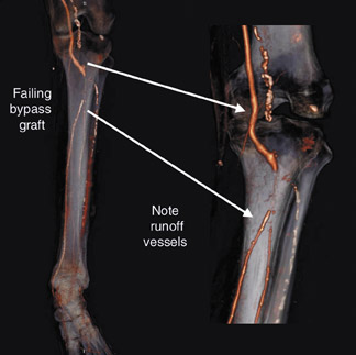
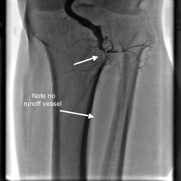
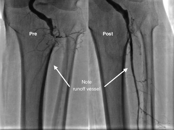
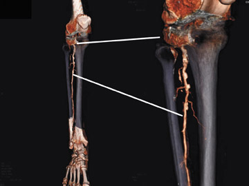
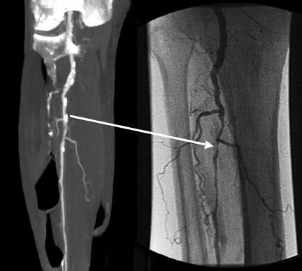
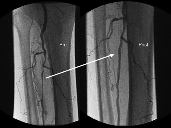
Rubin et al 21 reported the identification of 26 additional infrainguinal arterial segments utilizing CTA that were not identified with DSA because of the improved arterial opacification distal to an occluded segment. CTA-facilitated identification of "distal targets" could identify patients for tibial bypass or endovascular revascularization who otherwise may be offered only amputation. One of the most common reasons for an amputation recommendation is "no distal runoff target" (Figures 4B and C). Underscoring the need for simpler, accurate, noninvasive diagnostic imaging, we reported a series of 417 CLI patients of whom 67% had a primary amputation as their initial treatment without undergoing vascular evaluation. 26 Of these primary amputated patients, only 49% had an ankle-brachial index (ABI), and only 16% underwent diagnostic angiography. 26 This becomes important when considering the excellent >93% 6-month limb salvage rates reported by Laird et al 27 utilizing the excimer laser (Spectranetics Corporation, Colorado Springs, CO) in the multicentered LACI trial. Similar 12-month results were reproduced by our group in the "LACI-equivalent" report. 28
Clinical therapeutic benefits of PV-CTA
We have experienced the following understated clinical benefits with PV-CTA. Illustrative cases will be provided.
1. Vascular access (VA) planning. An underestimated benefit of PV-CTA is the ability to assist in VA planning, which is much more complex in peripheral vascular interventions (PVI) than in coronary interventions. The PVD patient often has limited VA with poor or no femoral pulses, complex femoral grafts, heavy femoral calcification, significant groin scarring, and previously deployed stents, which often impinge upon the CFA (Figure 5). For these reasons, VA complications are high in PVI, with a reported incidence of 8% to 16%. 30 We have drastically decreased our incidence of VA complications since utilizing preprocedural PV-CTA planning. The choice of alternate VA directed by PV-CTA (ie, brachial and radial artery) has significantly expanded our use of endovascular interventions, decreased vascular access complications, and improved outcomes in patients who otherwise would have required major open surgical revascularization procedures or suffered VA complications during PVI.
2. Clinical diagnostic accuracy. The combination of a thorough clinical examination and outpatient PV-CTA has facilitated an accurate preprocedural diagnosis in even our most complex PVD patients. The diagnosis is often rendered in the office in minutes, not hours and days. Onsite PV-CTA and conventional angiographic validation studies have been performed at our facility in all vascular territories, revealing >95% sensitivity and specificity and further providing confidence in our preprocedural diagnosis and PVI planning. 4,5
3. Therapeutic treatment planning and case performance. Likewise, PV-CTA facilitates reliable procedure planning, even including revascularization options and device planning. Lesion morphology characterizations now assist in treatment planning and periprocedural decision making. Calcified lesions are treated differently from soft lesions (ie, laser versus PTA versus cryoplasty versus stenting), and thrombus-containing lesions are treated differently from intimal hyperplastic lesions (ie, laser versus plaque excision versus DPD use versus mechanical thrombectomy versus lysis) (Figure 5, D through F). Peripheral vascular intervention cases are often complex in performance and decision making and are therefore longer in duration and have a much higher risk for overall complications than coronary interventions. The number of periprocedural PVI "surprises" we encounter today is dramatically diminished when utilizing PV-CTA; therefore, decreasing our overall therapeutic PVI procedural times, radiation exposure, and contrast utilization would facilitate overall improved outcomes.
We have found CTA of infrapopliteal arteries to be particularly helpful in periprocedural planning in patients with CLI. Utilizing our CLI protocol and a second lower extremity scan, we regularly identify patent distal infrapopliteal and pedal vessels-"distal targets"-that have not been previously imaged by angiography. We advocate at least our CLI-CTA on all patients before amputation. The identification of these "CTA-identified" but "not angiography-identified" vessels have significant therapeutic implications for the CLI patient, facilitating appropriate PVI or surgical bypass planning strategies (Figure 4, B through D).
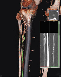
4. Bypass graft and stent surveillance. Infrainguinal bypass grafts and PVIs have a 20% to 30% secondary intervention rate to achieve an acceptable 1- to 2-year patency, which underscores the need for noninvasive postprocedural surveillance. 30 Angiography in patients after bypass grafting can be complex and is associated with increased VA complications. We avoid "sticking" a previous bypass graft for PVI unless no other access is available, and we use CTA in all patients with bypass grafts to decrease complications. Willmann et al 30 reported 98% sensitivity and specificity in comparing DSA versus 4-channel CTA in 85 bypass grafts. In our experience, CTA has allowed more accurate diagnosis and planning for graft reintervention than has DU and has decreased complications by facilitating vascular access planning during PVI. Unlike coronary stents, which are small and not well imaged on CTA, peripheral stents are much larger, and current software technology allows CTA to be an excellent tool for stent interrogation for stent fracture, ISR, and edge dissections (Figure 6).
5. Decreased overall complications. Peripheral vascular CTA has almost totally eliminated the need for and risk of diagnostic carotid angiography with its small but definite risk of stroke. Contrast-induced nephropathy (CIN) has been reported in 14% of PCI patients, with high mortalities and morbidities. 31 The incidence and impact of CIN in PVI is unknown and is very likely underestimated. The PVD patient is generally 10 to 15 years older than the PCI patient, and, invariably, PVD patients require multiple procedures and secondary reinterventions.
The risk of CIN is highly associated with intra-arterial contrast exposure but less associated with intravenous (IV) contrast exposure as used during PV-CTA. With proper patient selection and when outpatient oral and IV hydration protocols are used, CT-induced CIN should be exceedingly rare. We have developed PV-CTA protocols to decrease IV contrast volume exposure to 70 mL while retaining imaging quali- 4,5 Consequently, our periprocedural PVI intra-arterial contrast volume and CIN incidence has dramatically decreased when combining a preprocedural CTA-contrast planning strategy with clinically validated periprocedural IV hydration protocols during PVI.
6. Incidental vascular and nonvascular disease. CTA imaging acquisition also retains traditional CT nonvascular tissue capabilities. Occult neoplasms, severe degenerative arthropathies, spinal stenosis, and cholelithiasis are examples of additional clinical information with therapeutic implications that are frequently encountered during PV-CTA. Severe RAS, AAAS, and iliac, visceral, and popliteal artery aneurysms are also frequently encountered during abdominal CTA with runoff for the assessment of lower extremity occlusive disease. Likewise, vascular occlusive disease is often encountered in patients being investigated for aneurysmal disease. The identification and treatment of unknown RAS during the treatment of elderly patients with CLI is commonplace and facilitates the overall therapeutic care of this high-risk patient population who often require bilateral procedures with frequent reinterventions and, thus, receive significant contrast exposures.
Conclusion
Peripheral vascular CTA is an emerging tool in not only the diagnostic but also the overall therapeutic treatment of PVD. Further experience will expose the many understated clinical benefits of this technology in the comprehensive management of PVD. Peripheral vascular CTA has become an equally important tool in our PVD "therapeutic tool box" as wires, balloons, lasers, and stents.
Acknowledgment
The authors wish to thank Kelly Tilbe, NCMA, NCPT, for her assistance with technical manuscript preparation.
REFERENCES
- Yost ML. Peripheral arterial disease underestimated, undertreated. Volume II: A significant opportunity. Atlanta, GA: The Sage Group; 2004.
- NationalHospital Discharge Survey:Annual Summary with Detailed Diagnosis and Procedure Data.Data from the National Hospital Discharge Survey.Series 13. 1983-2000. Bethesda, MD: U.S. Department of Health and Human Services. National Center for Health Statistics; 2003.
- Faxon DP, Creager MA, Smith SC Jr, et al. Atherosclerotic Vascular Disease Conference: Executive summary: Atherosclerotic Vascular Disease Conference proceeding for healthcare professionals from a special writing group of the American Heart Association. Circulation. 2004;109:2595-2604.
- Allie DE, Hebert CJ, Lirtzman MD, et al. 64-channel multidetector computed tomography in non-cardiac vascular disease. Abstract presented at: Cardiovascular Imaging 2005: 33rd Annual Meeting & Scientific Sessions of the North American Society for Cardiac Imaging; October 11, 2005; Amelia Island, FL.
- Allie DE. Non-cardiac computed tomography angiography: Role of 64-channel CTA in the diagnosis and treatment of patients with critical limb ischemia. Abstract presented at: International Congress XXI, Endovascular Interventions; February 13, 2007; Scottsdale, AZ.
- Pan XM, Saloner D, Reilly LM, et al. Assessment of carotid artery stenosis by ultrasonography, conventional angiography, and magnetic resonance angiography: Correlation with ex vivo measurement of plaque stenosis. J Vasc Surg. 1995;21:82-89.
- Elgersma OE, Buijs PC, Wüst AF, et al. Maximum internal carotid arterial stenosis: Assessment with rotational angiography versus conventional intraarterial digital subtraction angiography. Radiology. 1999;213:777-783.
- Lorenz MW, von Kegler S, Steinmetz H, et al. Carotid intima-media thickening indicates a higher vascular risk across a wide age range: Prospective data from the Carotid Atherosclerosis Progression Study (CAPS). Stroke. 2006;37:87-92.
- Kaufmann TJ, Kallmes DF. Utility of MRA and CTA in the evaluation of carotid occlusive disease. Semin Vasc Surg. 2005;18:75-82.
- Leclerc X, Godefroy O, Lucas C, et al. Internal carotid arterial stenosis: CT angiography with volume rendering. Radiology. 1999;210:673-682.
- Link J, Brossmann J, Penselin V, et al. Common carotid artery bifurcation: Preliminary results of CT angiography and color-coded duplex sonography compared with digital subtraction angiography. AJR Am J Roentgenol. 1997;168:361-365.
- Hirai T, Korogi Y, Ono K, et al. Maximum stenosis of extracranial internal carotid artery: Effect of luminal morphology on stenosis measurement by using CT angiography and conventional DSA. Radiology. 2001; 221:802-809.
- Akers DL, Markowitz IA, Kerstein MD. The value of aortic arch study in the evaluation of cerebrovascular insufficiency. Am J Surg. 1987;154:230-232.
- Allie DE, Hebert CJ, Lirtzman MD, et al. Intraoperative innominate and common carotid intervention combined with carotid endarterectomy: A "true" endovascular surgical approach. J Endovasc There. 2004; 11:258-262.
- Beregi JP, Elkohen M, Deklunder G, et al. Helical CT angiography compared with arteriography in the detection of renal artery stenosis. AJR Am J Roentgenol. 1996;167:495-501.
- Galanski M, Porkop M, Chavan A, et al. Accuracy of CT angiography in the diagnosis of renal artery stenosis [in German]. Rofo. 1994;161:519-525.
- Shammas NW, Kapalis MJ, Dippel EJ, et al. Clinical and angiographic predictors of restenosis following renal artery stenting. J Invasive Cardiol. 2004; 16:10-13.
- AbuRahma AF, Stone PA, Bates MC, Welch CA. Angioplasty/stenting of the superior mesenteric artery and celiac trunk: Early and late outcomes. J Endovasc Ther. 2003;10:1046-1053.
- Silva JA, White CJ, Collins TJ, et al. Endovascular therapy for chronic mesenteric ischemia. J Am Coll Cardiol. 2006;47:944-950.
- Martin ML, Tay KH, Flak B, et al. Multidetector CT angiography of the aortoiliac system and lower extremities: A prospective comparison with digital subtraction angiography. AJR Am J Roentgenol. 2003;180:1085-1091.
- Rubin GD, Schmidt AJ, Sofilos MC, et al. Multi-detector row CT angiography of lower extremity arterial inflow and runoff: Initial experience. Radiology. 2001;221:146-158.
- Katz DS, Hon M. CT angiography of the lower extremities and aortoiliac system with a multi-detector row helical CT scanner: Promise of new opportunities fulfilled. Radiology. 2001;221:7-10.
- Fleischmann D, Hallett RL, Rubin GD. CT angiography of peripheral arterial disease. J Vasc Interv Radiol. 2006;17:3-26.
- Allie DE, Hebert CJ, Walker CM. Nitinol stent fractures in the SFA: The biomechanical forces exerted on the SFA provide a 'stiff' challenge to endovascular stenting. Endovasc Today. July/Aug 2004:1-8.
- Scheinert D, Scheinert S, Sax J, et al. Prevalence and clinical impact of stent fractures after femoropopliteal stenting. J Am Coll Cardiol. 2005;45:312-315.
- Allie DE, Hebert CJ, Lirtzman MD, et al. Critical limb ischemia: A global epidemic. A critical analysis of current treatment unmasks the clinical and economic costs of CLI. EuroIntervention. 2005;1(1):75-84.
- Laird JR, Zeller T, Gray BH; LACI Investigators. Limb salvage following laser-assisted angioplasty for critical limb ischemia: Results of the LACI multicenter trial. J Endovasc Ther. 2006;13:1-11.
- Allie DE, Hebert CJ, Walker CM. Excimer laser-assisted angioplasty in severe infrapopliteal disease and CLI: The CIS "LACI equivalent" experience. Vasc Dis Manage. 2004;1(1):14-
- Shammas NW, Lemke JH, Dippel EJ, et al. In-hospital complications of peripheral vascular interventions using unfractionated heparin as the primary anticoagulant. J Invasive Cardiol. 2003;15:242-246.
- Willmann JK, Mayer D, Banyai M, et al. Evaluation of peripheral arterial bypass grafts with multi-detector row CT angiography: Comparison with duplex US and digital subtraction angiography. Radiology. 2003; 229:465-474.
- McCullough PA, Wolyn R, Rocher LL, et al. Acute renal failure after coronary intervention: Incidence, risk factors, and relationship to mortality. Am J Med. 1997;103:368-375.
Related Articles
Citation
Peripheral vascular CTA: Emerging role of PV-CTA in the therapeutic management of PVD. Appl Radiol.
April 3, 2008