Cardiovascular Case Report: Coronary artery involvement in acute aortic dissection with pseudoaneurysm: Evaluation with cardiac
Images


Prepared by Jonathan D. Dodd, MB, David O'Donnel, MB, Ronan Killeen, MB, Ronan Ryan, MB, Peter Quigley, MB, and Martin Quinn, MB, St. Vincent's University Hospital, Elm Park, Dublin, Ireland.
Case History
A 73-year-old woman was admitted for the investigation of atypical, intermittent chest pain and dyspnea. Her chest pain came on suddenly, was not relieved by nitrates, and did not radiate. It continued to occur intermittently throughout her hospital stay but remained stable in severity. Her dyspnea was more gradual in onset, was mild initially, and became progressively worse over the course of her stay. Her surgical history was significant for an aortic valve replacement 20 years previously for aortic stenosis. She did not suffer from hypertension and was a nonsmoker. Her medications included anticoagulation for her prosthetic valve.
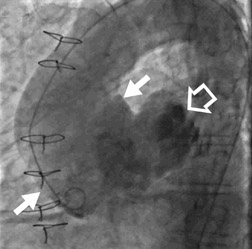
On examination, the patient had a mild tachycardia (100 bpm) and normal blood pressure. An ejection systolic murmur and click was auscultated over the aortic area. Her electrocardiogram, serum troponin, and chest radiograph were normal. Over the next 10 days she remained stable, but her intermittent chest pain persisted and her dyspnea progressed. Because of her continuing atypical chest pain, she was referred to invasive coronary angiography. The cardiologist was unable to engage the coronary arteries. An aortic angiogram was performed and the aortic lumen was found to be widely dilated (Figure 1). A large outpouching was noted arising from the posterior wall of the ascending aorta.
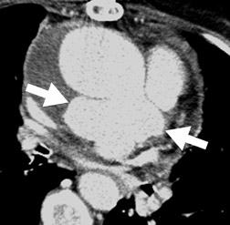
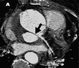
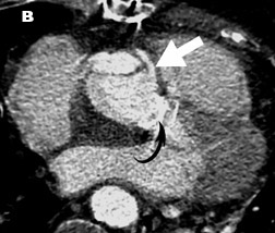
The patient was transferred urgently to the radiology department, where she underwent 64-slice cardiac CT (Siemens Sensation, Erlangen, Germany) to further evaluate the aorta and coronary arteries. Technical parameters included 375-msec rotation time, 0.75-mm slice thickness, 0.4-mm overlap, kVp 120, 850 mAs, pitch 0.2, 120 mL of Niopam 370 (Bracco UK, Ltd., High Wycombe, UK) (using the test bolus technique), and image reconstruction with retrospective electrocardiographic (ECG) gating. The images showed a Stanford type A aortic dissection. A portion of the dissection had partially ruptured through the posterior aortic wall to form a large pseudoaneurysm (Figure 2). The pseudoaneurysm was causing significant external compression on the right main pulmonary artery (Figure 3). The 64-slice cardiac CT images clearly showed extension of the dissection flap to involve the ostium of the left main (LM) coronary artery (Figure 4A). The right coronary artery ostia appeared uninvolved (Figure 4B). Based on the cardiac CT findings, surgical repair included an LM coronary reimplantation in addition to an aortic root and valve replacement. The patient made a full, immediate postoperative recovery.
Discussion
Recent developments in CT technology have led to its increasing application for acute chest pain. For our patient, it provided valuable preoperative information on the extension of the dissection flap to involve the LM coronary artery, as well as the presence of a large pseudo-aneurysm. Technically, if a dissection does not involve the coronary arteries, then the aortic graft repair is performed as a supracoronary procedure, and the native aortic shell becomes wrapped around the graft. If the coronary arteries are involved by the dissection, coronary graft reimplantation (Bentall procedure) is necessary. 1 Thus, in acute aortic dissection, coronary artery involvement plays a major role in the type of surgical procedure performed. Traditionally, the coronary arteries have not been evaluable with CT. The rapid motion of the heart resulted in excessive motion artifacts, precluding robust assessment of the coronary ostia in the context of aortic dissection. 2 Newer CT techniques involving data reconstruction with ECG-gating allow images to be reconstructed at a particular part of the R-R interval throughout scan acquisition. Usually, the best portion is at 60% to 70% of the R-R interval, resulting in "motionless" imaging of the coronary arteries. The images in the current case were reconstructed at 65% of the R-R interval, allowing excellent visualization of the proximal extent of the dissection to the LM coronary ostium.
The application of CT angiography for patients referred with chest pain is currently an area of intense research interest. Several studies have shown cardiac CT to have a negative predictive value of close to 100% in the exclusion of acute coronary syndrome. 3 Dedicated cardiac CT protocols are evolving to include a "triple-rule-out" assessment for aortic dissection and pulmonary embolism in addition to the coronary arteries, but these protocols lack thorough clinical evaluation. Early studies have shown that CT may provide excellent visualization of the coronary, pulmonary, and aortic territories. 4 Such technology developments are supporting recommendations that cardiac CT may play an important role in the investigation of patients presenting to the emergency department with acute chest pain. 5
Conclusion
Cardiac CT with retrospective ECG-gating allows accurate evaluation of coronary ostial involvement in acute aortic dissection.
References
- Erbel R, Alfonso F, Boileau C, et al. Diagnosis and management of aortic dissection. Eur Heart J. 2001;22:1642-1681.
- Batra P, Bigoni B, Manning J, et al. Pitfalls in the diagnosis of thoracic aortic dissection at CT angiography. RadioGraphics. 2000;20:309-320.
- Hoffmann U, Nagurney JT, Moselewski F, et al. Coronary multidetector computed tomography in the assessment of patients with acute chest pain. Circulation. 2006;114:2251-2260. Erratum in: Circulation. 2006; 114:e651.
- Johnson TR, Nikolaou K, Wintersperger BJ, et al. ECG-gated 64-MDCT angiography in the differential diagnosis of acute chest pain. AJR Am J Roentgenol. 2007;188:76-82.
- Stillman AE, Oudkerk M, Ackerman M, et al. Use of multidetector computed tomography for the assessment of acute chest pain: A consensus statement of the North American Society of Cardiac Imaging and the European Society of Cardiac Radiology. Eur Radiol. 2007;17:2196-2207.
Related Articles
Citation
Cardiovascular Case Report: Coronary artery involvement in acute aortic dissection with pseudoaneurysm: Evaluation with cardiac . Appl Radiol.
June 13, 2008