MRI Improves Staging of Patients with Small Cell Lung Cancer
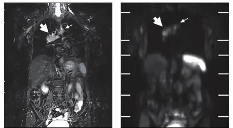
 A study published in the American Journal of Roentgenology (AJR) reports that MRI, with or without FDG PET co-registration, improves the staging of patients with small cell lung cancer (SCLC) compared to conventional staging tests.
“FDG PET/CT, whole-body MRI, and co-registered FDG PET/MRI outperformed conventional tests for various staging endpoints in patients with SCLC,” concluded first author Yoshiharu Ohno from the Fujita Health University School of Medicine in Japan. Whole-body MRI and FDG PET/MRI outperformed FDG PET/CT for T category and thus TNM stage, “indicating utility of MRI for assessing extent of local invasion in SCLC.”
Ohno and colleagues’ prospective study included 98 patients (64 men, 34 women; median age, 74 years) with SCLC who underwent conventional staging tests (brain MRI; neck, chest, and abdominopelvic CT; bone scintigraphy), FDG PET/CT, and FDG PET/MRI within 2 weeks before treatment. After MRI technologists performed co-registration via proprietary software (Canon Medical Systems), two nuclear medicine physicians and two chest radiologists independently reviewed the examinations in separate sessions.
In patients with SCLC, accuracy for T category was higher (p<.05) for whole-body MRI (94.9%) and FDG PET/MRI (94.9%) than for FDG PET/CT (85.7%). Meanwhile, TNM stage accuracy was higher (p<.05) for whole-body MRI (88.8%) and FDG PET/MRI (86.7%) than for FDG PET/CT (77.6%) and conventional staging tests (72.4%).
“These additional observations may relate to a superior role of MRI in assessing the extent of local soft tissue invasion by tumor, as has been observed in settings other than SCLC,” the authors said.
Related Articles
Citation
MRI Improves Staging of Patients with Small Cell Lung Cancer. Appl Radiol.
December 10, 2021