Infant Brain Atlases Unveiled by UNC Researchers
 Pew-Thian Yap, PhD, professor in the University of North Carolina Department of Radiology, and colleagues in the department and the Biomedical Research Imaging Center (BRIC) have created a new collection of month-by-month infant brain atlas (IBA) based on MR imaging that capture fine details of the early developing brain across both space and time.
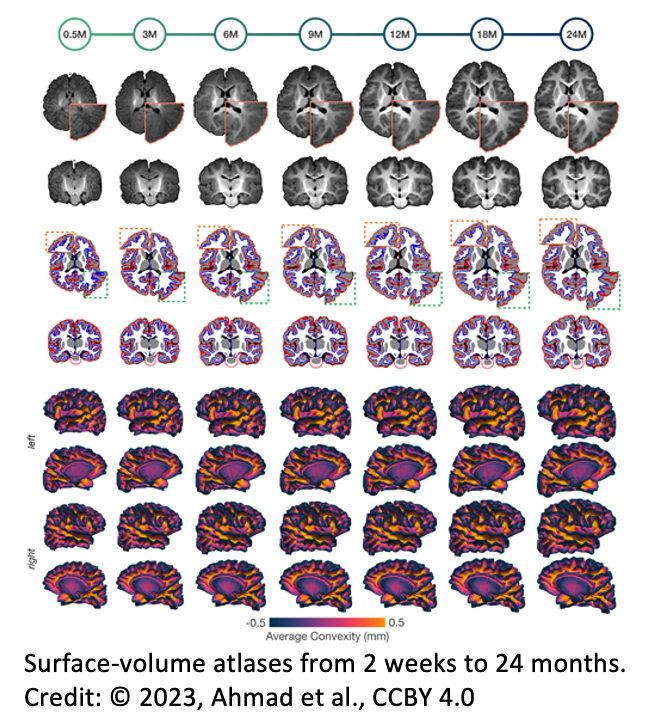
In this work, published in the journal Nature Methods, the scientists created a set of month-specific surface-volume longitudinal brain atlases of infants from 2 weeks to 2 years of age. Sahar Ahmad, PhD, research instructor of radiology, was lead author on the paper.
“Brain atlases are key to understanding neurodevelopment through the lenses of cellular composition, neural pathways, and functional organization,” Dr Yap said. “The human brain atlases created by our team depict the early development phase of postnatal neurodevelopment. Our atlases will be a resource valuable to brain scientists in unraveling key normative and aberrant traits of, arguably, the most important phase of human brain development.”
Throughout the first two years of life, the human brain undergoes a whole range of cellular processes that drive the rapid growth of the infant brain. It is during this period that the brain changes structurally and reorganizes its neural circuits. When development goes awry, it can have detrimental effects on the quality of life, including heightened risk for autism, schizophrenia, and ADHD. By using the IBA, researchers are able to capture heterogeneous changes in brain structure, cortical geometry, and tissue contrast.
A widely used measurement for the cortical geometry is called cortical thickness. In terms of neuroanatomy, cortical thickness refers to the thickness of the brain’s gray matter — the outermost layer of nerve cell tissue. The IBA that was developed by Yap and colleagues shows that cortical thickness increases rapidly during the first year and more slowly in the second year.
The atlases also revealed that cortices in the temporal, parietal, and prefrontal regions of the brain are thicker than the primary visual and sensorimotor cortices. This is consistent with the finding that the higher-order functions of the infant brain – such as attention, working memory, inhibition, and problem-solving – mature more slowly than the areas of the brain that are responsible for the visual, motor, and sensory functions.
Overall, the surface-volume consistent IBA accurately captures the infant growth trajectories and does so with rich anatomical details. These atlases recorded the monthly changes in the normally developing brains’ size, shape, and cortical geometry as well as their tissue contrast, volume, and microstructural characteristics from 2 weeks to 2 years of age.
“We hope that these atlases will become a common coordinate framework to facilitate the discovery of new insights into developmental processes underpinning child cognition and social behavior,” Dr Yap said.
Related Articles
Citation
Infant Brain Atlases Unveiled by UNC Researchers. Appl Radiol.
January 30, 2023