Incidental atypical sclerosing papilloma within a radial scar on digital breast tomosynthesis
Images



CASE SUMMARY
A 39-year-old female presented to the breast imaging center with a three-week history of a new, non-tender, palpable right breast lump at 12:00. The patient was pre-menopausal and had no personal or family history of cancer. Baseline bilateral diagnostic mammography and ultrasound were performed for further evaluation.
IMAGING FINDINGS
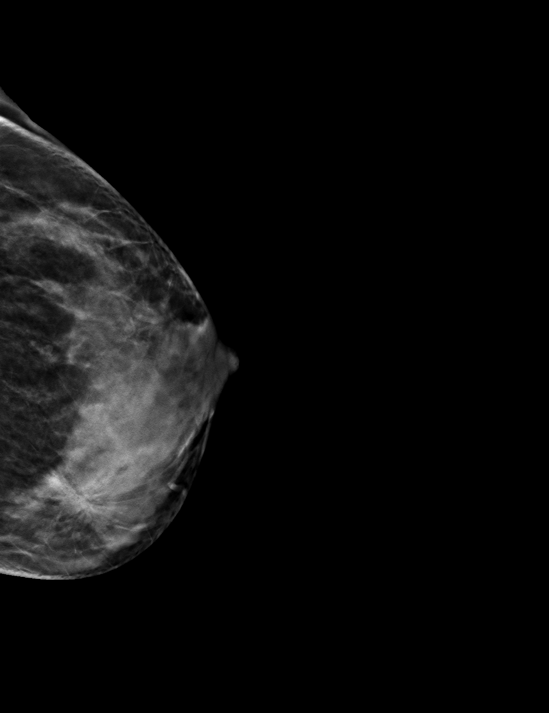
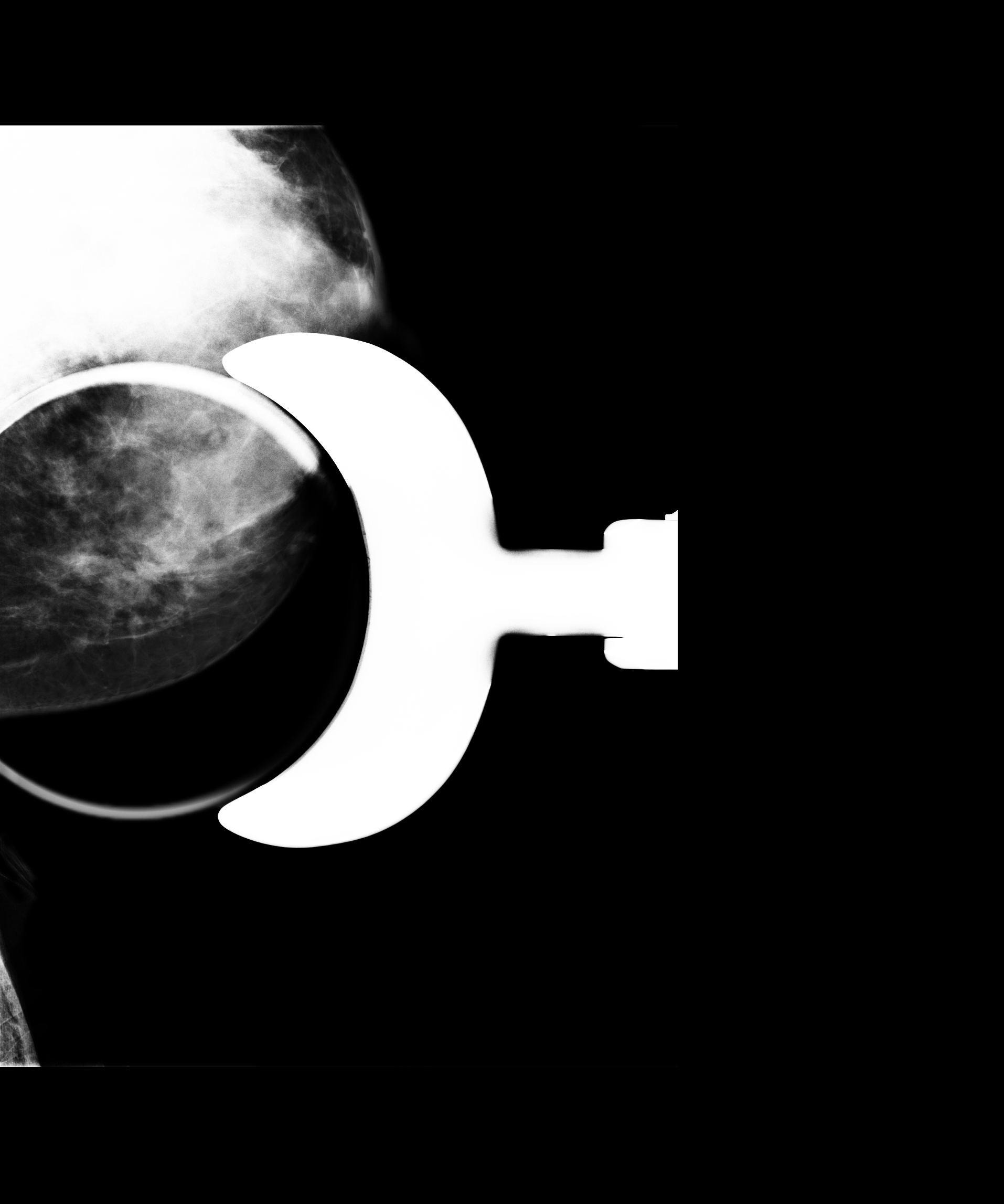
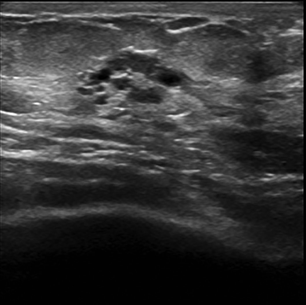
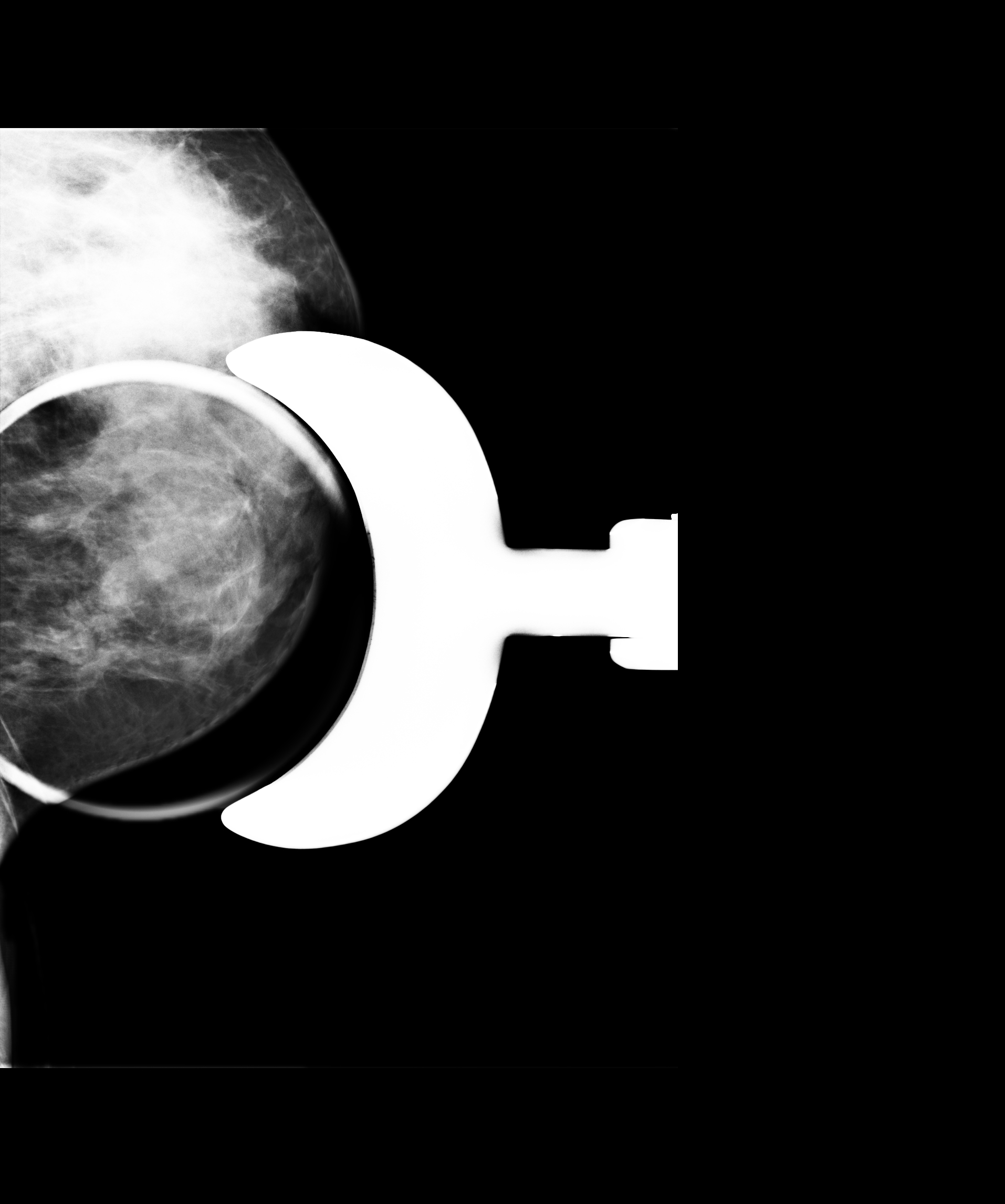
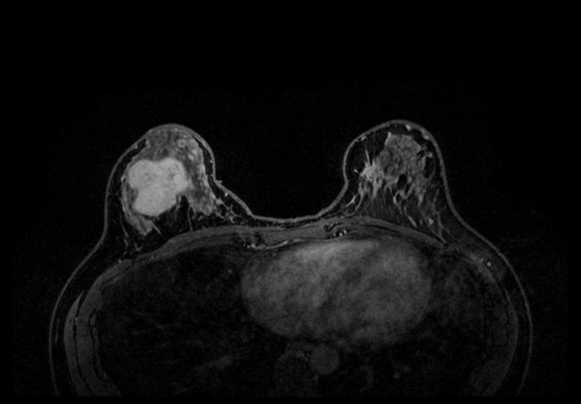
Digital breast tomosynthesis (DBT) images of both breasts were performed. Mammography and ultrasound of the right breast revealed a heterogeneous circumscribed solid mass with moderate associated color flow measuring 4.8 cm (Figure 1). Incidentally, in the left breast, a prominent area of architectural distortion was identified on mammography, best seen medially on the craniocaudad view (Figure 2). Additional 2-D spot compression views demonstrated persistent architectural distortion in the lower inner quadrant of the left breast (Figure 3). Targeted left breast ultrasound revealed focal fibrocystic changes on the left (Figure 4). Bilateral contrast-enhanced breast MRI showed a large homogeneously enhancing mass in the right breast and a highly suspicious 2.2 cm irregular mass with spiculated margins and associated washout in the lower inner quadrant of the left breast (Figure 5). Surgical excisional biopsy confirmed a benign fibroadenoma in the right breast, and an atypical sclerosing papilloma in the setting of a radial scar/complex sclerosing lesion in the left breast.
DIAGNOSIS
Atypical sclerosing papilloma within a radial scar/complex sclerosing lesion
DISCUSSION
Radial scars (RS), also known as complex sclerosing lesions (CSL), are benign breast lesions. By convention, RS corresponds to a lesion measuring < 1 cm, while a CSL corresponds to a lesion > 1cm of the same histology.1 They are often clinically concerning as they share radiographic and histologic features that are very similar to carcinoma. The clinical importance of diagnosing a radial scar is threefold: 1) RS is difficult to distinguish from carcinoma, 2) foci of intraductal or invasive carcinoma can at times be found within or adjacent to a RS, and 3) RS may potentially act as a marker for increased risk for invasive breast cancer or ductal carcinoma in situ.2
Mammographic and clinical features that can suggest a diagnosis of RS include an asymmetry or area of architectural distortion (AD) with areas of central translucency, with the absence of a discrete, palpable central mass or skin changes. Classically, RS is described as having a spiculated appearance with a “black star” effect in which the spicules are surrounded by an absence of a solid tumor mass.2 Recent literature has demonstrated that DBT might offer the advantage of improved detection of AD over 2-D mammography.3,4 Furthermore, a cancer detection rate of 21% is reported when AD is discovered on DBT. DBT identified AD that would otherwise remain undetected by traditional mammography.3
Ultrasound is another potential modality that can augment the effort to differentiate radial scars from carcinomatous findings. It is important to note that there is no one defining sonographic feature that distinguishes RS from stellate carcinomas. In general, RS has the appearance of a focal area of heterogeneous echogenicity, with or without posterior acoustic shadowing, and with an absence of a discernable mass, or of a solid mass with or without an irregular border. Importantly, RS are often more conspicuous on ultrasound than on mammography.5 On the other hand, MRI findings of RS are typically composed of a stellate lesion without mass effect in precontrast images, and often exhibit benign kinetic behavior with absent or mild contrast uptake on post-contrast imaging.2
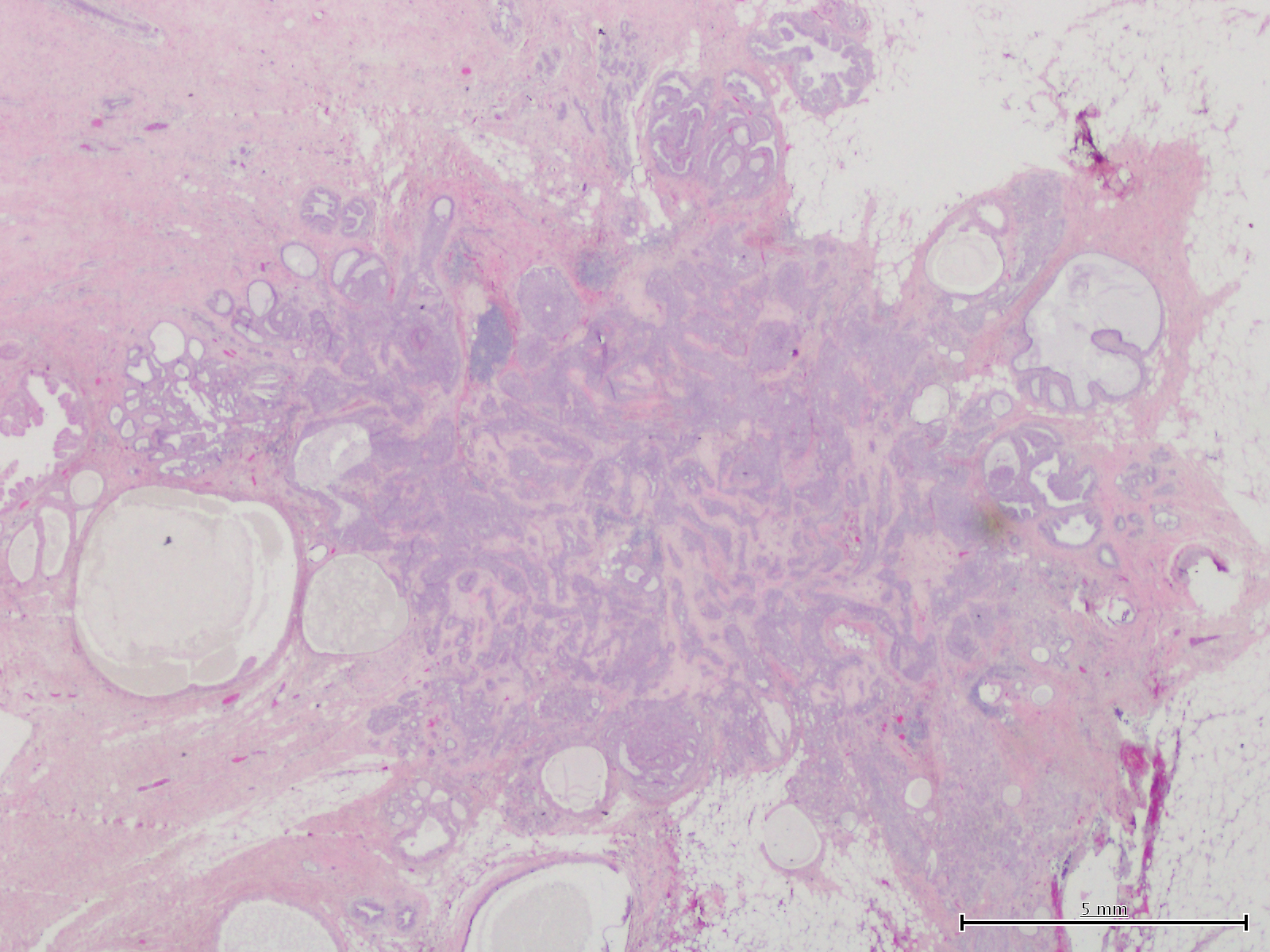
Histologically, RS is described as a radially symmetric entity consisting of a fibro-elastotic core from which ducts and lobules radiate, and which may contain epithelial tubules (Figure 6). The typical histologic pattern of RS is what results in the frequent mimicry of breast cancer on mammography.6
A diagnosis of RS was traditionally diagnosed via surgical excisional breast biopsy. This strategy was sub-optimal since many of the findings represented carcinomas that subsequently required a second surgery to completely resect the newly diagnosed malignancy.7 A diagnosis of RS can now be attained via percutaneous core needle biopsy. However, when a potential cancer is suspected, an accurate diagnosis of RS without malignancy may require more core tissue samples as compared to biopsy of a carcinoma. Core needle biopsy in the setting of adequately sampled RS may eliminate the need for surgical biopsy.8
Still, malignancy is often underestimated even in the setting of percutaneous biopsy-proven RS due to sampling and diagnostic error attributed to the difficulty in differentiating RS from carcinoma. Furthermore, mammographic and sonographic findings of RS are not always predictive of malignancy at surgery. As such, it is for the most part still recommended that RS found on percutaneous biopsy be surgically excised in order to definitely exclude the presence of malignancy.9
CONCLUSION
Radial scars/complex sclerosing lesions are benign lesions of the breast that pose a diagnostic challenge due to their radiologic resemblance to breast cancer. The most common mammographic appearance is a spiculated area of parenchyma with a “black star” effect and a lack of a central tumor mass. Digital breast tomosynthesis with ultrasound may offer some benefit over traditional mammography in terms of detection of architectural distortion and, thus, radial scars. Although multiple core tissue samples obtained by percutaneous core needle biopsy may improve diagnostic yield and mitigate the need for a subsequent surgical biopsy, the current recommendation for management of most RS found on percutaneous biopsy is surgical excision.
REFERENCES
- Page D, Anderson T. Radial scars and complex sclerosing lesions. Diagnostic histopathology of the breast. 1st ed. Edinburgh, Scotland: Churchill Livingstone; 1987. Chapter 9, Radial scars and complex sclerosing lesions; p. 89-103.
- Linda, A, Zuiani C, Londero V, Cedolini C, Girometti R, Bazzocchi M. Magnetic resonance imaging of radial sclerosing lesions (radial scars) of the breast. E J Radiol. 2012; 81:3201-3207.
- Partyka L, Lourenco AP, Mainiero MB. Detection of mammographically occult architectural distortion on digital breast tomosynthesis screening: initial clinical experience. AJR Am J Roentgenol. 2014;203:216-222.
- Yang TL, Liang HL, Chou CP, Huang JS, Pan HB. The adjunctive digital breast tomosynthesis in diagnosis of breast cancer. BioMed Res Int. 2013;2013:597253.
- Lee E, Wylie E, Metcalf C. Ultrasound imaging features of radial scars of the breast. Australian Radiology. 2007;51:240-245.
- Cawson, J, Nickson C, Evans J, Kavanagh AM. Variation in mammographic appearance between projections of small breast cancers compared with radial scars. J Med Im and Rad Onc. 2010;54:415-420.
- Farshid G, Rush G. Assessment of 142 stellate lesions with imaging features suggestive of radial scar discovered during population-based screening for breast cancer. Am J Surg Pathol. 2004;28:1626-1631.
- Brenner RJ, Jackman RJ, Parker SH, Philpotts L, Deutch BM, Lechner MC, Lehrer D, Sylvan P, Hunt R, Adler SJ, Forcier N. Percutaneous core needle biopsy of radial scars of the breast: when is excision necessary? AJR Am J Roentgenol. 2002; 179:1179-1184.
- Linda A, Zuiani C, Furlan A, Londero V, Girometti R, Machin P, Bazzocchi. Radial scars without atypia diagnosed at imaging-guided needle biopsy: how often is associated malignancy found at subsequent surgical excision, and do mammography and sonography predict which lesions are malignant? AJR Am J Roentgenol. 2010;194:1146-1151.
Citation
Fitzpatrick SS, KA, Lebeau, LG, MH MWB. Incidental atypical sclerosing papilloma within a radial scar on digital breast tomosynthesis. Appl Radiol. 2017;(7):44-47.
July 1, 2017