Hologic Adds 3D Breast Ultrasound to SuperSonic MACH 40
 Hologic, Inc. has announced that 3D ultrasound imaging is now available on the SuperSonic™ MACH™ 40 ultrasound system. Clinicians can now access high-resolution B-mode and ShearWave™ PLUS elastography 3D volumes, which are designed to enhance diagnostic certainty.
With the 3D volumetric data acquired on the SuperSonic MACH 40 system, clinicians can see areas of interest from a new perspective. Breast tissue can be visualized in any scanning plane of the 3D volume, including coronal or C-plane. MultiSlice display allows these 3D volumes to be viewed slice-by-slice, while MultiPlanar display virtually reconstructs the slices in any orientation using the acquired and stored volume. The system’s volumetric transducer allows users to easily and rapidly acquire these 3D images with no resolution loss, regardless of where a lesion is located within the breast.
The additional diagnostic details provided by 3D imaging may assist clinicians in the workup of difficult lesions, including in patients with dense breast tissue.[1] Furthermore, pairing 3D imaging with the system’s ShearWave PLUS elastography may also contribute to more accurate tumor size estimation[2] and clear margin definition in pre-operative settings. In addition, the combination may play a role in monitoring and evaluating breast cancer patients during and after neoadjuvant chemotherapy.[3]
“At Hologic, we relentlessly strive to advance the early detection of breast cancer,” said Jennifer Meade, Hologic’s Division President, Breast and Skeletal Health Solutions. “With each innovation across the breast care continuum, we’re moving toward greater certainty for our customers and enabling them to provide better outcomes for their patients. The addition of 3D breast ultrasound imaging to the SuperSonic MACH 40 system is yet another example of the steps we’re taking to transform the daily experience of breast radiologists and sonographers with solutions designed to increase efficiency and accuracy, while also helping to improve diagnostic confidence.”
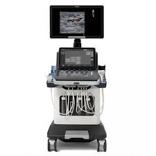
Thanks to its exclusive UltraFast™ imaging technology, the SuperSonic MACH 40 system has an image capture capacity of up to 20,000 frames per second,[4] which ensures smooth images with reduced speckle and improved lesion conspicuity for enhanced diagnostic confidence. This technology powers the system’s exceptional image quality and many of its innovative imaging modes.
The system’s control panel features the revolutionary SonicPad™ touchpad, which makes the user experience more intuitive and helps streamline workflow by reducing user movement and overall examination time.
[1] Berg WA, Blume JD, Cormack JB, et al. Combined screening with ultrasound and mammography vs. mammography alone in women at elevated risk of breast cancer [published correction appears in JAMA. 2010 Apr 21;303(15):1482]. JAMA. 2008;299(18):2151-2163. doi:10.1001/jama.299.18.2151
[2] Accuracy of tumor size measurement: Comparison of B-mode ultrasound, strain elastography, and 2D and 3D shear wave elastography with histopathological lesion size. Farrokh A, Maass N, Treu L, et al. Acta Radiol. 2018;60(4):451-458. doi: 10.1177/0284185118787354. | Shear-wave elastography contributes to accurate tumour size estimation when assessing small breast cancers. Mullen R et al. Clin Radiol. 2014 Dec;69(12):1259-63.
[3] Feasibility of Imaging and Treatment Monitoring of Breast Lesions with Three-Dimensional Shear Wave Elastography. Athanasiou A, Latorre-Ossa H, Criton A, Tardivon A, Gennisson JL, Tanter M. Ultraschall Med. 2015 Mar 5. | Comparison of strain and shear-wave ultrasonic elastography in predicting the pathological response to neoadjuvant chemotherapy in breast cancers. Ma Y et al. Eur Radiol. 2017 Jun;27(6):2282-2291. | Shear-Wave Elastography for the Detection of Residual Breast Cancer After Neoadjuvant Chemotherapy. Lee SH et al. Ann Surg Oncol. 2015 Dec;22 Suppl 3:S376-84.
[4] Ultrafast Ultrasound Imaging, by Jeremy Bercoff (Published: August 23rd 2011 DOI: 10.5772/19729)