Hemangiopericytoma of the Third Ventricle
Images
CASE SUMMARY
A 70-year-old with a history of atrial fibrillation and prostate cancer, and thalassemia had a third ventricular mass incidentally detected on a brain MRI.
IMAGING FINDINGS
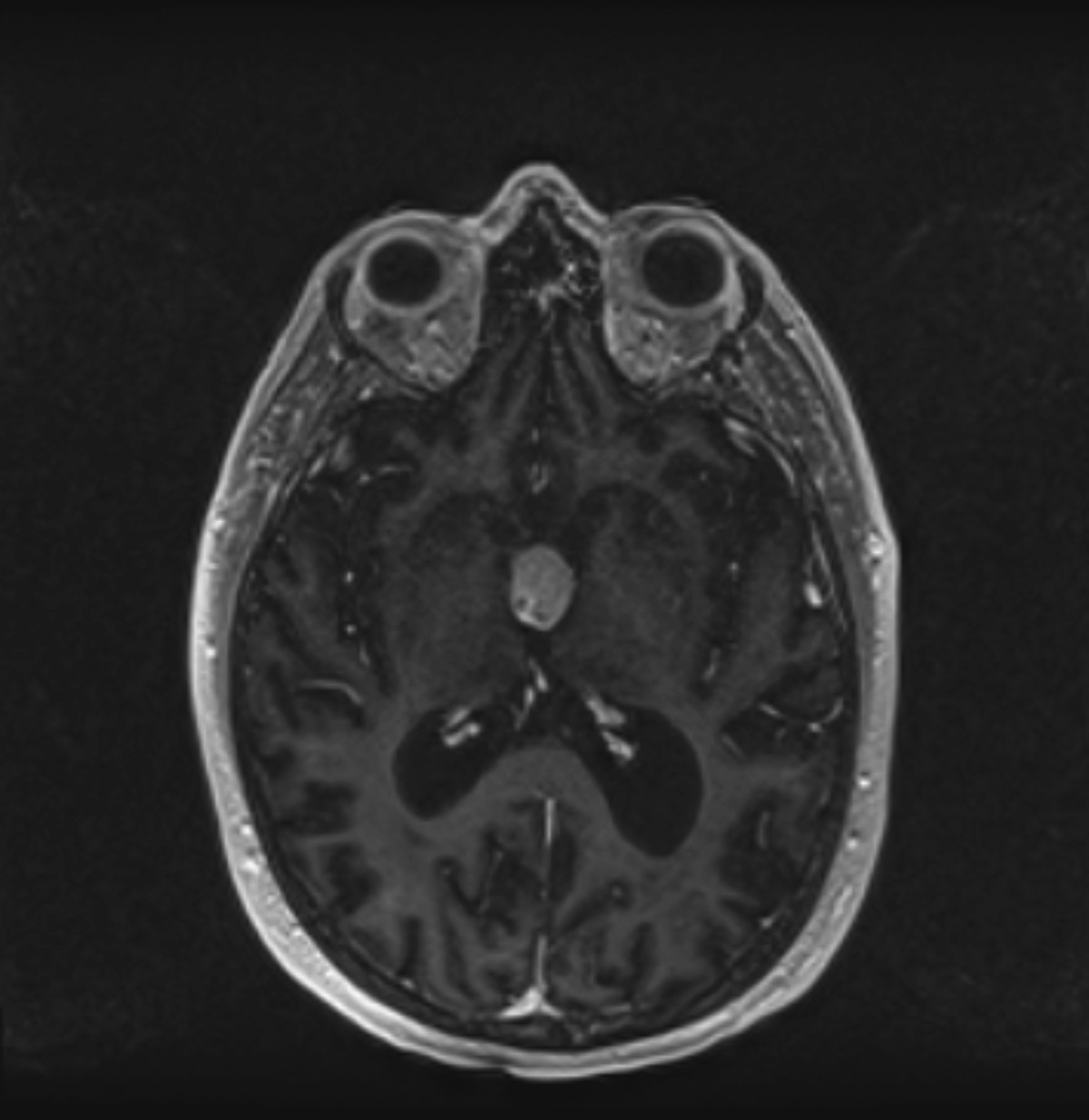
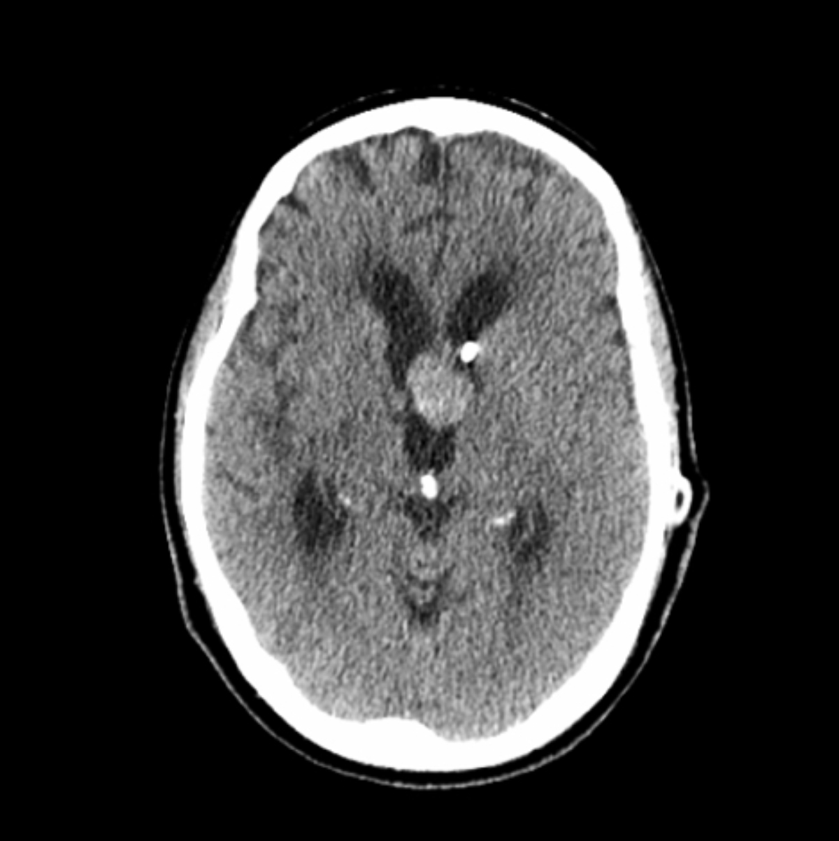
A contrast-enhanced T1 image showed an enhancing mass located in the anterior third ventricle near the foramen of Monro (Figure 1). Endoscopic biopsy of the mass demonstrated histologic findings consistent with hemangiopericytoma. CT 5 months later demonstrated that the mass had grown (Figure 2).
DIAGNOSIS
Hemangiopericytoma of the third ventricle
DISCUSSION
Hemangiopericytoma (HPC) is a central nervous system tumor that arises from the pericytes of meningeal capillaries.1 These tumors are more likely to occur in males, most commonly in the fourth or fifth decade.1 Signs and symptoms at presentation may include headache, blurred vision, nausea, vomiting, and photophobia.4 Intraventricular HPC is rare and most often found in the lateral ventricles. HPC in the third ventricle are exceedingly rare, with only one previous case found in our literature search.2
Intraventricular HPC may arise from pericytes within the tela choroidea or choroid plexus.3 There are many pathological and imaging similarities between HPC and angioblastic/anaplastic meningioma. Histologically, HPC shows a staghorn vascular pattern of spindly cells and are reactive to vimentin, but not to epithelial membrane antigen, unlike meningioma, which is positive for both substances.5 One of the most characteristic features of HPC is a dense reticulin network composed of individual cells.4
On imaging, intracranial HPC tends to occur supratentorially along meningeal surfaces, with most occurring in parasagittally. They are usually oval, have lobulated margins, are dense on CT, and have avid contrast enhancement on CT and MRI.5 They may show vascular flow voids, as in our case, and cystic areas. Peripheral HPC lacks the “dural tail” sign typical of meningioma.5,6 Differential diagnostic considerations for intraventricular masses include include colloid cyst, central neurocytoma, ependymoma, subependymal giant cell astrocytoma and intraventricular meningioma.
Although third ventricular HPC is extremely rare, their consideration can be important. Their noncontrast appearance and location can mimic those of a colloid cyst.7 Colloid cyst, however, is a non-neoplastic lesion. Biopsy of an HPC may be more likely to hemorrhage than other third ventricular lesions, given its hypervascularity.5 In our case, the patient developed postoperative intraventricular and subarachnoid hemorrhage (Figure 2). HPC also has a propensity to recur and metastasize through the CNS and beyond. Therefore, gross total surgical resection is the goal.5 Postoperative adjuvant radiotherapy can lower risk of recurrence.7
CONCLUSION
This case demonstrates an extremely rare intraventricular hemangiopericytoma of the third ventricle. While the imaging features of these lesions can be similar to those of other intraventricular masses, consideration of HPC is important, given their vascularity and risk of hemorrhage, as well as their similar noncontrast appearance to the colloid cyst.
REFERENCES
- Thakur P, Sharma M, Gupta M, Chatterjee D, Fotedar V. Anaplastic cerebral hemangiopericytoma: Rare variant of a rare disease. Clin Cancer Invest. J. 2015; 4: 277-279.
- Abrahams JM, Forman MS, Lavi E, Goldberg H, Flamm ES. Hemangiopericytoma of the third ventricle. Case report. J Neurosurg. 1999;90 (2): 359-362.
- Towner, JE, Johnson MD, Li YM. Intraventricular Hemangiopericytoma: A Case Report and Literature Review. World Neurosurg. 89: 728-728.
- Abdollahi A, Abdollahpouri R, Tavangar S-M. Meningeal Hemangiopericytoma in 33-Year-Old Female; a Case Report. Iranian J Pathol. 2016;11(3): 281-285.
- Pang H, Yao Z, Ren Y, Liu G, Zhang J, Feng X. Morphologic patterns and imaging features of intracranial hemangiopericytomas: a retrospective analysis. OncoTargets and Therapy. 2015; 8: 2169-2178.
- Sotoudeh H, Yazdi HR. A review on dural tail sign. World Journal of Radiology. 2010;2(5): 188-192.
- O’Neill AH, Gragnaniello C, Lai LT. Natural history of incidental colloid cysts of the third ventricle: A systematic review. J Clin Neuroscience. 2018; 53: 122-126.
Citation
S K, K K.Hemangiopericytoma of the Third Ventricle. Appl Radiol. 2020; (5):47-48.
September 1, 2020