Fourth update on CTA of 24 novel coronary stents
Descriptions of the appearance and characteristics of coronary stents on coronary computed tomography angiography (CTA) have expanded. The fourth update and in vitro evaluation of 24 new novel stent types by radiologists at the University Hospital of Cologne in Germany have been published in the September issue of Acta Radiologica.
These evaluations of stent lumen visualization are intended to help radiologists interpreting examinations of patients after stent placement and helping physicians decide which stents are suitable for assessment by coronary CTA.

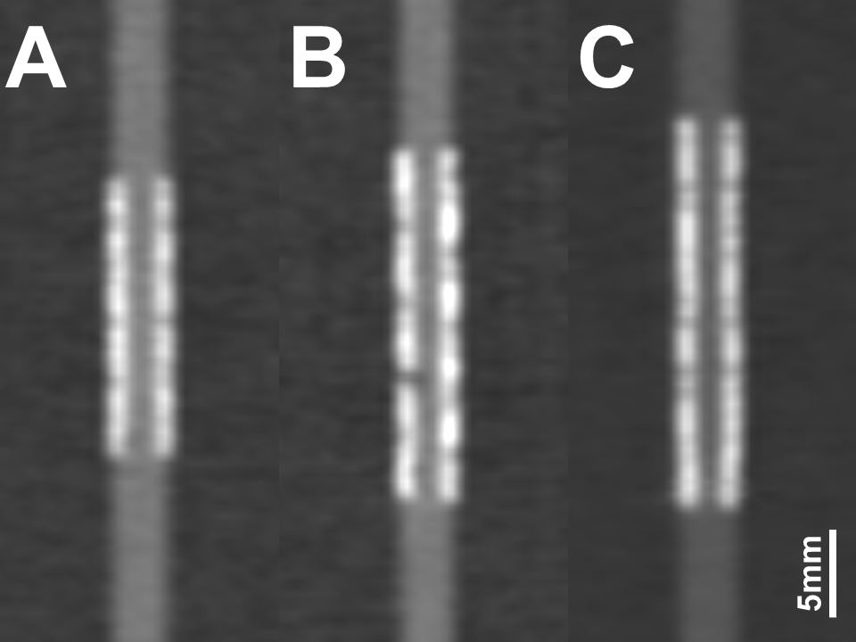
Example images of 3 different coronary stents
manufactured from cobalt-chromium (A),
stainless-steel (B) and platinum-chromium (C)
Stent-related artifacts impair in-stent lumen visibility. This can impede accurate reassessment of the vessel lumen when monitoring for the presence of in-stent restenosis, the renarrowing and/or blockage of a coronary artery after stent treatment. Stent architecture, stent strut thickness, and/or the material composition of a stent influence the related artifacts on CT images.
Updating their prior catalogues, the authors evaluated stents have become clinically available since 2014, utilizing current CT acquisition and reconstruction technologies1,2,3. They used a 128-row spectral detector CT scanner with a simulated electrocardiogram signal with a heart rate of 60 beats per minute to evaluate 17 cobalt-chromium stents, six stainless-steel stents, and one platinum-chromium stent in a coronary phantom. Using standard coronary CTA parameters and stent-specific reconstruction algorithms they quantified image quality for each stent with respect to in-stent attenuation alteration and visible lumen diameter.
Principal investigator Tilman Hickethier, MD, and colleagues reported that six stents showed a lumen visibility of 56% to 60%, and 14 stents a lumen visibility of 45% to 55%, with the lowest recorded lumen visibility of 40% in one stent. Thirteen stents showed deviations of the in-stent lumen attenuation of up to 10% and 11 stents caused moderate alterations of the measured densities inside of the stent lumen up to 20%. The authors wrote that none of the 24 stents showed severe deviations of in-stent attenuation.
Cobalt-chromium has replaced stainless-steel as the most frequent manufacturing medium for stents over the past 10 years, according to the authors. However, differences in mean visible diameters were identical. The single platinum-chromium stent had a smaller visible diameter (1.23 mm compared to 1.52 mm) and higher attenuation alterations (35.04 Hounsfield Units compared to 21.25 HU), but also produced acceptable images.
“All of these novel stents appear eligible for evaluation by coronary CTA,” the authors concluded.
REFERENCES
- Maintz D, Seifarth H, Raupach R, et al. 64-slice multi-detector coronary CT angiography: in vitro evaluation of 68 different stents. Eur Radiol. 2006;16(4):818-826.
- Maintz D, Burg MC, Seifarth H, et al. Update on multi-detector coronary CT angiography of coronary stents: in vitro evaluation of 29 different stent types with dual-source . Eur Radiol. 2009;19(1)42-49.
- Gassenmaier T, Petri N, Allmendinger T, et al. Next generation coronary CT angiography: in vitro evaluation of 27 coronary stents. Eur Radiol. 2014;24(11):2953-2961..
- Hickethier T, Wenning J, Doerner J, et al. Fourth update on CT angiography of coronary stents: in vitro evaluation of 24 novel stent types. Acta Radiol. 2018;59(9):1060-1065.
Citation
Fourth update on CTA of 24 novel coronary stents. Appl Radiol.
December 5, 2018