Philips Announces New AI Applications for Cardiovascular Ultrasound Systems
Images

Royal Philips announced its next-generation AI-enabled cardiovascular ultrasound platform to help speed up cardiac ultrasound analysis with proven AI technology. Integrated into the company’s EPIQ CVx and Affiniti CVx ultrasound systems, the new FDA 510(k) cleared AI applications significantly advance cardiovascular imaging and diagnosis, automating measurements and speeding workflows to increase productivity.
Heart failure is a rapidly growing health issue that affects an estimated 64 million people worldwide. It's also associated with high mortality, and poor quality of life and is a substantial burden on healthcare systems globally. As the most common and least invasive way to check the structure and function of the heart, cardiovascular ultrasound has played a key role in diagnosing cardiac disease earlier.
“As clinical cases get more complex and patient volumes increase, we read hundreds of echocardiography exams daily with thousands of data points,” said Roberto Lang, MD, Director of the Noninvasive Cardiac Imaging Lab, University of Chicago Medicine, USA. “With the integration of AI into echocardiography solutions, we can now automate some of the steps to support clinicians' decision-making, allowing them to detect, diagnose, and monitor various cardiac conditions with greater confidence and efficiency.”
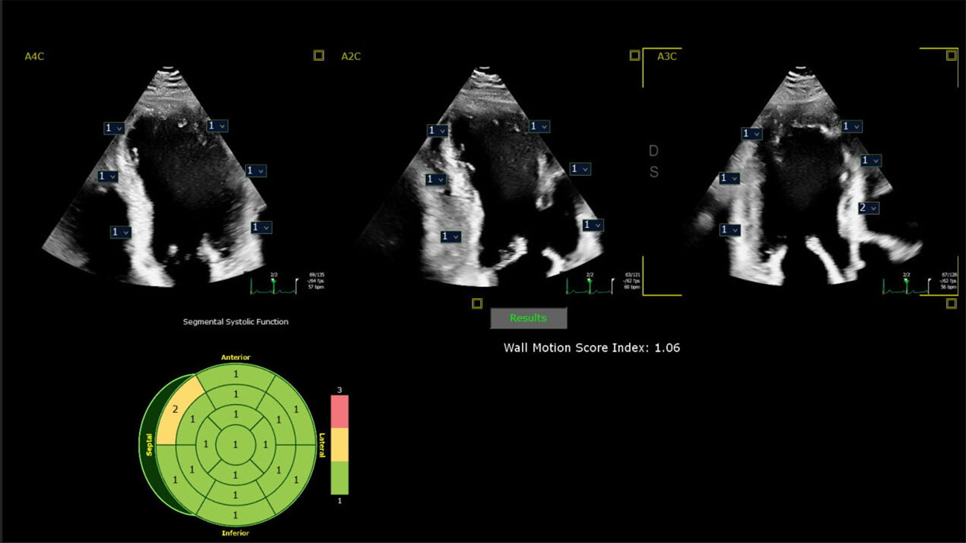
Dr Lang will join other clinicians to share the results of a new scientific abstract being presented at the American Society of Echocardiography (ASE2024) annual meeting, June 14-16, demonstrating how first-of-kind AI algorithms co-developed with Philips provide highly accurate detection of regional wall motion abnormalities (RWMA) on echocardiography. RWMAs can be an independent indicator of adverse cardiovascular events and death in patients with cardiovascular diseases like myocardial infarction (MI) and congenital heart disease. Automated machine learning-based assessment of RWMA has the potential to improve the efficiency of all readers. “An advantage of AI methods over conventional visual analysis is that it can be performed in seconds, providing rapid and accurate information to help augment expert reads by quickly highlighting areas of concern for RWMA, improving the ease and efficiency of interpretation,” Lang added.
“By harnessing the power of AI into our echocardiography solutions, we empower clinicians with enhanced diagnostic capabilities, ultimately improving patient care and outcomes in the management of coronary and valvular disease, while enhancing overall efficiency in cardiac practice,” said David Handler, VP and General Manager for Global Cardiovascular Ultrasound at Philips. “For patients, this means consistent image interpretation which can lead to fewer re-scans, shorter and more effective interventional procedures, and potentially faster recovery times.”
Trained on anonymized patient data sets from real-life clinical environments, the AI features integrated across Philips cardiovascular ultrasound systems help improve the quality and reproducibility of cardiac imaging and enhance operator and departmental efficiency. These include FDA-cleared and CE-marked software solutions from DiA Imaging Analysis. Together, these AI features automate how users interpret ultrasound images, so clinicians with varying levels of ultrasound experience can automatically analyze images with increased speed, efficiency, and accuracy in real-time. In addition to integrating these latest advanced AI features into the company’s cardiovascular ultrasound systems, Philips’ new X11 4t Mini TEE ultrasound transducer, introduced earlier this year, is now fully compatible with the EPIQ CVx and Affiniti CVx systems.