Low-Count Quantitative SPECT Shows Promise for Personalizing Cancer Treatment
Images
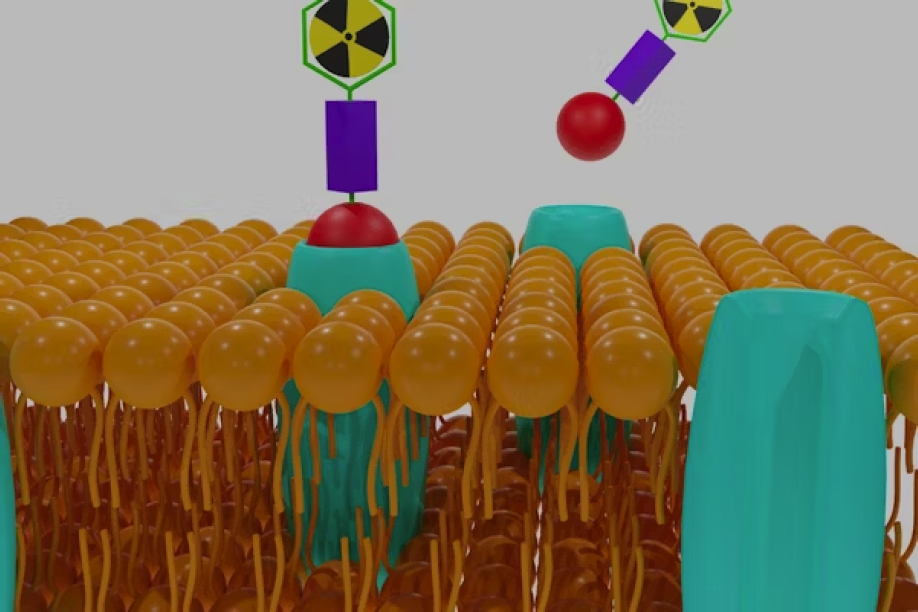
A group of investigators led by Abhinav Jha, assistant professor of biomedical engineering in the McKelvey School of Engineering and of radiology at Mallinckrodt Institute of Radiology in the School of Medicine at Washington University in St. Louis, developed a new imaging method for personalized cancer treatment. Recently published results from a computer-based clinical trial indicate that the method may be reproducible in humans and merits additional clinical testing.
In a study published in the Journal of Nuclear Medicine, Jha and collaborators reported the results of a computer-based imaging trial that evaluated the effectiveness of a specialized single-photon emission computed tomography (LC-QSPECT) imaging method to estimate doses of alpha-particle-emitting radiopharmaceutical therapy (a-RPT). The trial simulated 280 virtual patients with bone-metastatic prostate cancer. Results showed that LC-QSPECT provided reliable dose estimates across different scanner setups, with high accuracy, repeatability and reproducibility across scanners, outperforming conventional methods. These results indicate that LC-QSPECT holds promise to enhance the safety and effectiveness of personalized treatment, encouraging further clinical evaluations.
The authors concluded that the “projection-domain LC-QSPECT method yielded both high reproducibility and high accuracy across multiple scanner–collimator configurations and high test–retest repeatability on the task of estimating dose for a-RPTs.” They further noted that LC-QSPECT outperformed the conventional OSEM method on the estimation task.
Related Articles
Citation
Low-Count Quantitative SPECT Shows Promise for Personalizing Cancer Treatment. Appl Radiol.
June 20, 2024