How AI is Revolutionizing Opportunistic Detection in CT
Images

Opportunistic findings are those that are unexpected or incidental and discovered on medical imaging studies performed for other reasons. Artificial intelligence (AI) can help detect these small, and sometimes subtle, findings, leading to earlier treatment of potentially serious conditions.
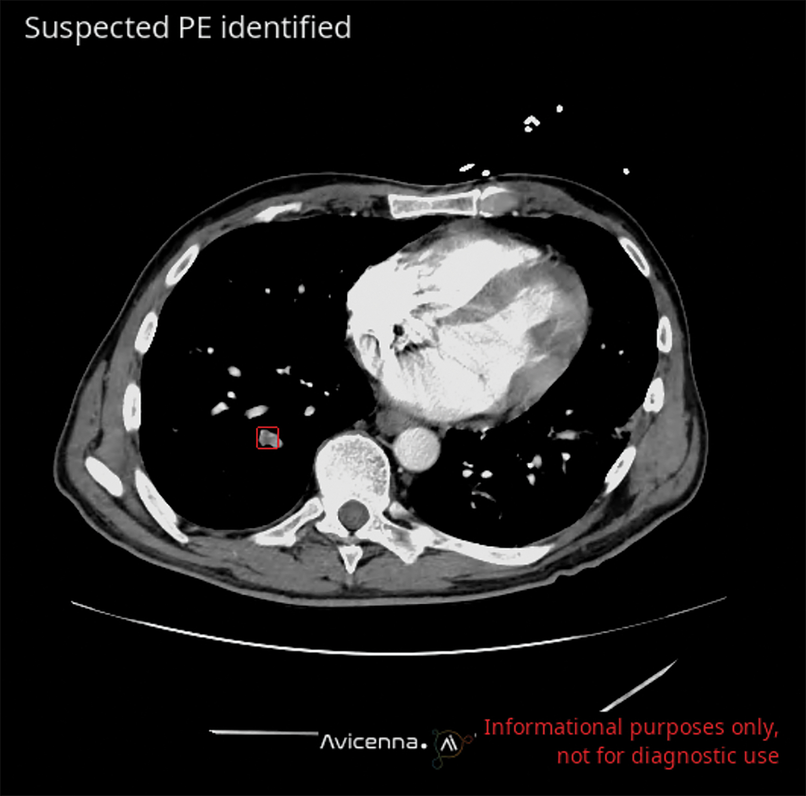
Opportunistic detection of pulmonary embolism (PE) is one example among many. The frequency of incidental detection of PE among oncology patients is approximately 3.4%.1 Up to 45% of PEs are missed by radiologists when “off-search” for the exam’s primary purpose.2 Undetected PE is associated with poorer outcomes; thus, prompt management is essential.
Oncology studies may be reviewed anywhere from hours to days after they have been acquired. This is particularly true of late, with the growing backlog of examinations associated with rising case volumes and staffing limitations.
Capable AI can work in the background, leveraging computer vision as an always-on assistant, unfettered by indication-related expectations, and ensuring that opportunistic findings are promptly identified, called to the radiologists’ attention, and reported.
Incidental detection of disease on common examinations like CTs can also have a significant impact on health. For example, using AI to scrutinize CT scans acquired for other reasons can help identify unsuspected vertebral compression fractures (VCFs). These fractures can be difficult to detect, especially in patients who are not experiencing severe pain. AI-based screening of CT scans can lead to earlier treatment, preventing complete vertebral collapse, and improving patient quality of life.3,4
AI algorithms can also be used with CT to quantify visceral fat when evaluating for metabolic syndrome; to assess muscle bulk and density for sarcopenia diagnosis; to quantify liver fat in assessing hepatic steatosis; and to quantify aortic and coronary calcium for cardiovascular risk.5,7
These algorithms provide reproducible and reliable measurements to assess the hidden condition and help personalize patient management. Early studies also show their potential to predict treatment response and future adverse events.8,9
The American College of Radiology’s Incidental Findings Committee, supports opportunistic identification and quantification of coronary calcium on routine CT scans of the chest as well as CT performed at low dose for lung cancer screening.10
Although these applications of AI have the potential to improve patient outcomes and increase efficiency, they also have drawbacks. It is true that some findings can be beneficial, but others may be clinically insignificant and yet lead to additional imaging, testing, and higher costs, as well as psychological distress, for patients. Convenient methods of informing the patient about the opportunistic finding and ensuring adequate follow-up are necessary for the appropriate use of such capabilities.
At present, only a limited number of AI-fueled applications are available for screening, particularly for diseases with low incidence. But more are expected in the coming years, and they may be able to weigh patient risk factors and contribute to disease prediction and prevention.
References
Citation
S Q. How AI is Revolutionizing Opportunistic Detection in CT. Appl Radiol. 2023;(4):22-23.
June 23, 2023