BrainLab, FujiFilm Collaborate on Intraoperative Neurosurgery Solution
Images
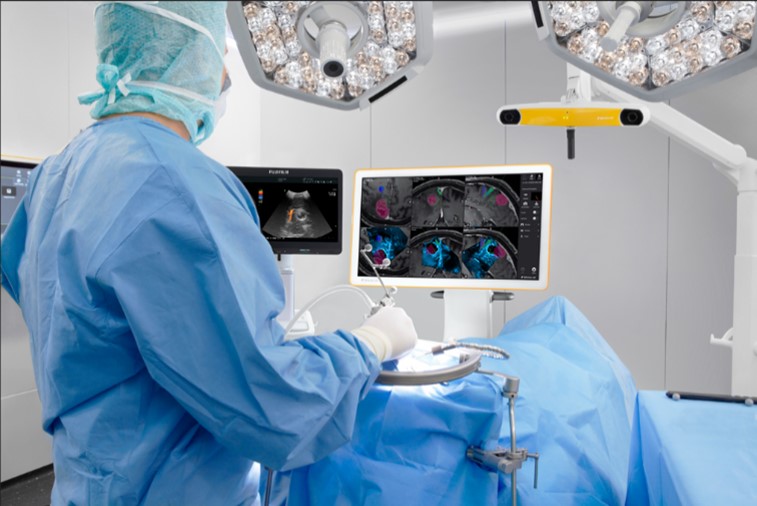
FUJIFILM Healthcare Americas Corporation and BrainLab announced that Brainlab will be the exclusive U.S. distributor of ARIETTA Precision Ultrasound for neurosurgery applications to be utilized with Brainlab’s surgical navigation systems. ARIETTA Precision, in conjunction with a Brainlab surgical navigation system, becomes a powerful intraoperative neurosurgery solution, delivering advanced guidance and image quality for critical surgical decisions. Together, the systems serve as a deeply integrated end-to-end solution providing comprehensive intraoperative information leveraging the best of both worlds in imaging and navigation. The new offering is an attractive expansion, poised to enhance surgical workflows for the over 2,000 neurosurgery departments across the US.
Brainlab will distribute the ARIETTA Precision as a standalone product alongside and soon with its Ultrasound Navigation software. This combined solution is pending FDA clearance and expected to be available in the US in mid-2024. The combination of these technologies provides neurosurgeons with real-time insights through live intraoperative ultrasound (IOUS) during neurosurgical procedures. The high quality, continuously updatable ultrasound images deliver a dynamic layer of confidence throughout the entire procedure, helping neurosurgeons achieve their goal of maximum safe tumor or lesion resection, even if brain shift occurs.
Studies show that navigation greatly improves the surgeon’s ability to approach, assess and operate on brain tumors, but may lose its accuracy as the surgery progresses and brain shift occurs. IOUS has proved to serve as a solution by providing real-time patient information that can be used to update navigation images and provide the neurosurgeon with real-time information on the resection progress.
Digital integration between Brainlab surgical navigation solutions and Fujifilm’s ARIETTA Precision, will offer streamlined system setup for cranial imaging and “plug-n-play” workflows with pre-calibrated and sterilizable neurosurgery transducers, eliminating the need for user calibration in the sterile field. Additionally, the combined technologies provide neurosurgeons with automatic view layouts, including the ability to superimpose intraoperative 3D ultrasound scans onto the preop dataset for a more comprehensive overview of the resection cavity. With this technology integration, neurosurgeons can positively impact their practice, their facility and most importantly, their patients.
“Fujifilm has been a leader in neurosurgical ultrasound for decades and is committed to bringing to market systems and probes that help neurosurgeons make critical surgical decisions,” said Hideyuki Honda, vice president, ultrasound solutions, FUJIFILM Healthcare Americas. “The ARIETTA Precision was specifically designed for surgeons, and we believe the deep technology integration of Brainlab’s sophisticated software platform with the ARIETTA Precision will not only provide neurosurgeons with the technology they need for critical decision making but will enhance patient care across the nation.”
Surgeons can easily visualize any misalignment between an intraoperatively acquired 3D ultrasound scan and a preoperative MRI with Brainlab Ultrasound Navigation software. Furthermore, with Elements ‘Ultrasound Snap to MRI’ (also known as Brainlab Elements Image Fusion) software, Brainlab enables automated rigid fusion between the preoperative MRI image and the intraoperative ultrasound image, allowing surgeons to update the registration and navigate on updated patient anatomy. R&D projects are already underway that will take the technology beyond rigid to elastic fusion, leveraging ultrasound images with virtual iMRI software to adapt and update.
“Fujifilm’s ARIETTA Precision ultrasound is an important addition to our neurosurgery portfolio,” said Sean Clark, president, Brainlab Inc. “Brainlab will leverage our market-leading advanced software capabilities to transform the ARIETTA Precision into a 3D image ‘digitizer,’ providing updates on patient anatomy throughout surgery.”