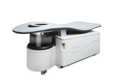
QT Imaging Gets FDA Nod to Calculate Fibroglandular Volume of the Breast
 The US FDA has given 510(k) clearance to QT Imaging to calculate the fibroglandular volume (FGV) of the breast and the ratio of FGV to total breast volume (TGV). This ratio can contribute to an assessment of risk for breast cancer and changes in this ratio can be used to measure the efficacy of medication when treating or preventing breast cancer.
Unlike traditional breast imaging modalities, the company's QTscan does not require radiation, injection, or compression, and is highly accurate (+ 0.2%), allowing earlier and more frequent monitoring for women undergoing non-surgical breast cancer treatments such as adjuvant chemotherapy, radiation therapy, cryotherapy, and hormone or selective hormone receptor modulation treatments. No other ultrasound-based breast imaging modality is cleared by the FDA to quantify fibroglandular volume.
"The ability to determine a therapeutic clinical response using a quantitative volumetric method is crucial for effective and timely treatment of breast cancer and for patients at high risk for developing breast cancer who are receiving hormonal therapy. The FGV tool will allow this assessment to be made early and in follow-up to maximize treatment benefit, which is an exciting development for breast care patients," said Dr. Elaine Iuanow, a Breast Imaging specialist and medical consultant for QT Imaging.
"We are excited to expand the tools available using low frequency transmitted ultrasound volography to serve women, especially those with dense breasts, providing an imaging option that is safe, comfortable, and effective," said Dr. John Klock, CEO, Chief Medical Officer and Founder of QT Imaging, Inc.
Specifically, the QT Scanner 2000 Model A is for use as an ultrasonic imaging system to provide reflection-mode and transmission-mode images of a patient's breast. The QT Scanner 2000 Model A software also calculates the breast fibroglandular tissue volume (FGV) value and the ratio of FGV to total breast volume (TBV) value as determined from reflection-mode and transmission-mode ultrasound images of a patient's breast. The device is not intended to be used as a replacement for screening mammography.