Unusual Imaging Finding in FDG-PET may be Omicron Infection
 The Society of Nuclear Medicine and Molecular Imaging (SNMMI) Task Force have recently observed an unusual imaging pattern on FDG PET/CT scans or FDG PET/MR scans that may be due to COVID-19 infection.
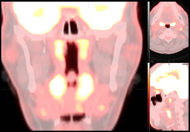
Unlike the FDG PET/CT (fluorodeoxyglucose (FDG)-positron emission tomography (PET) scan pattern seen with infections from previous strains of COVID-19, with principal involvement of the lungs, this new array of findings is primarily centered in the upper aerodigestive tract and cervical lymph nodes. Specifically, there is prominent, symmetric FDG uptake throughout the nasopharynx, oropharynx and tonsils with or without associated FDG-avid cervical lymphadenopathy, particularly in the suprahyoid neck. Based on what we know about the Omicron variant, it is conceivable that this pattern, whenever correlated with COVID-19 infection, is a result of the presently dominant Omicron strain.
The Task Force recommends that this pattern should be taken into consideration at the time of FDG PET/CT interpretations and the possibility of infection with Omicron variant of COVID-19 should be entertained in the differential diagnosis. Since this pattern can by no means be diagnostic of COVID-19 infection, the Task Force makes the following recommendations.
- Check the patient records to see if there is a recent positive COVID-19 test.
- Determine if the patient is at higher risk of COVID-19 infection based on current symptoms or due to recent exposure or travel. If so, a recommendation can be made to test for COVID-19 in the appropriate setting.
- Compare with prior FDG PET/CT examinations and the patient’s history to determine if this represents chronic inflammatory/reactive process or stable malignancy such as lymphoma.
- Various differential diagnostic possibilities should be considered if this pattern is new or if there is interval progression including, but not limited to, infection with COVID-19, other viruses such as Epstein-Barr virus, malignancy and bacterial infections.
- This pattern may also be seen in children and younger adults but should be interpreted cautiously in view of normal increased activity that can be physiologic. Correlation with history and symptoms and comparison to prior examinations is recommended.
Related Articles
Citation
. Unusual Imaging Finding in FDG-PET may be Omicron Infection. Appl Radiol.
January 25, 2022