The Ubiquity of AI at RSNA 2019
Images

At the 2019 Radiological Society of North America’s scientific exhibition in Chicago in December, nearly every company highlighted new artificial intelligence (AI) initiatives and enhancements to their diagnostic imaging systems. From AI developments in machine learning (ML) to deep learning (DL) technologies, these advanced systems are being designed to effectively address clinical conditions, while simplifying physician workflows and departmental throughput.
Following is a recap of some of the most exciting AI-based tech that could be make an appearance in medical imaging departments and clinics in the months ahead:
After entering the U.S. market in 2018, United Imaging Healthcare (UIH) announced several FDA-pending AI capabilities across multiple modalities with DELTA, a DL enhancement to its uCT 7 series computed tomography system; HYPER Deep Learning Reconstruction in the routine PET/CT workflow of its uMI 550 system; and AI-Assisted Compressed Sensing (ACS) full-coverage image acceleration for its uMR 780 system.
“The Delta algorithm can be applied to lower-dose datasets in order to recreate something that is similar to a higher-dose dataset. This has a big application for lowering dose, which is obviously on the hearts and minds of everybody who uses CT,” said Jeffrey M. Bundy, PhD, Chief Executive Officer of UIH.
United Imaging also announced a new strategic partnership with BAMF Health, which is achieving AI-enabled precision medicine through molecular imaging and theranostics. Powered by UIH’s proprietary AI platform, BAMF Health clinics will diagnose and deliver personalized treatment to patients with cancer, while leveraging United Imaging’s PET/CT and PET/MR technology.
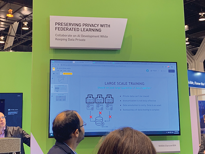
Gaining access to the huge volumes of data required to train AI models while protecting patient privacy is a key issue for healthcare facilities. NVIDIA is addressing this challenge with NVIDIA Clara Federated Learning (FL), which uses distributed training across multiple hospitals to develop robust AI models without sharing personal data.
iCAD showcased Profound AI for digital breast tomosynthesis (DBT), or 3D mammography, the first and only FDA-cleared AI solution to support breast cancer detection in DBT.
ProFound AI helps increase cancer detection rates while also reducing false positives and reading time to enhance clinical performance. According to a recent study, ProFound AI is clinically proven to increase sensitivity by 8%, reduce false positives by 7%, and slash radiologist reading time by 52%.
“With ProFound AI for DBT, radiologists are still in the driver’s seat – they’re just getting help with prioritizing which lesions to look at, which essentially cuts reading time in half,” said Jeff Hoffmeister, MD, MSEE, vice president and medical director of iCAD. “It’s especially helpful on challenging cases where a radiologist might need extra insight.”
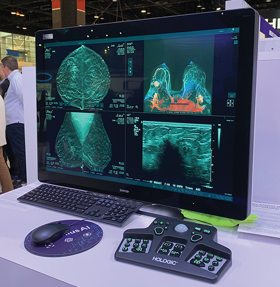
Hologic debuted the 3DQuorum Imaging Technology, powered by Genius AI, a breakthrough AI-powered algorithm to expedite mammography exam reading time without compromising image quality, sensitivity, or accuracy.
3DQuorum uses analytics enhanced by Genius AI to uniquely reconstruct high-resolution 3D data to produce 6 mm “SmartSlices.” The company says this expedites reading time by reducing the number of images for radiologists to review without compromising image quality, sensitivity, or accuracy. These analytics identify clinically relevant regions of interest and preserve important features during reconstruction of the SmartSlices.
Canon Medical Systems USA, Inc., is bringing AI to routine imaging with Advanced intelligent Clear IQ Engine (AiCE) Deep Learning Reconstruction, an innovative approach to CT reconstruction that uses DL to distinguish true signal from noise to deliver sharp, clear, and distinct images at fast speeds. Trained with vast amounts of high-quality image data, the FDA-pending AiCE provides enhanced anatomical resolution across the central nervous, respiratory, cardiac, and musculoskeletal systems.
This technology is already cleared by the FDA for use on the Aquilion ONE/GENESIS Edition and Aquilion Precision CT systems. It is pending FDA 510(k) clearance for use on the Aquilion ONE/PRISM Edition and the Aquilion Prime SP CT systems, as well as on the Vantage Galan 3T MR and Vantage Orian 1.5T MR systems.
Siemens Healthineers expanded its AI-Rad Companion family with two AI-based software assistants designed to free radiologists from routine activities during MRI examinations.
The AI-Rad Companion Brain MR for Morphometry Analysis automatically segments the brain in MRI images, measures brain volume, and marks volume deviations in result tables used by neurologists for diagnosis and treatment. AI-Rad Companion Prostate MR for Biopsy Support also automatically segments the prostate on MRI images and enables radiologists to mark lesions, facilitating targeted prostate biopsies.
GE Healthcare introduced several new digital applications built on Edison, the company’s digital intelligence platform, which was unveiled at RSNA 2018. The first, AIR Recon DL, is a new FDA-pending MR image reconstruction technology that delivers TrueFidelity Images. It’s designed to deliver greater image accuracy by leveraging advanced neural networks, or DL algorithms trained from a database of more than 10,000 images, to identify and remove artifacts, allowing users to optimize images and improve clinical diagnostics.
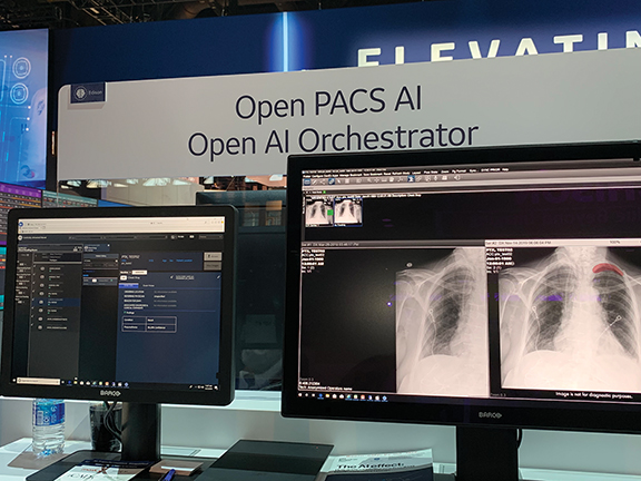
The new Edison Open AI Orchestrator manages imaging workflows. It simplifies implementation, deployment, support, and scaling of multiple AI applications, including those from partners iCAD and MaxQ, to seamlessly integrate clinical applications into the radiology PACS reading workflow. With this, Edison Open AI Orchestrator can help reduce the complexity of multiple systems and algorithms working together, potentially leading to fewer errors.
Edison Open AI Orchestrator seamlessly integrates multiple AI applications into the radiology PACS reading workflow.
FUJIFILM Medical Systems U.S.A., Inc. demonstrated how the FDA-pending FDR AQRO mini mobile DR system can be used in the ER with AI-powered recognition algorithms that could help physicians more clearly identify suspicious findings. AI recognition will search and map images to help identify pathologies such as pneumothorax.
Fujifilm’s FDX Console, also pending FDA clearance, will leverage AI in the OR with its ability to highlight foreign objects, such as surgical gauze, in images acquired during or post-surgery. AI for positioning navigation includes a camera built into the collimator of a Fujifilm X-ray unit to ensure proper patient positioning prior to exposure.
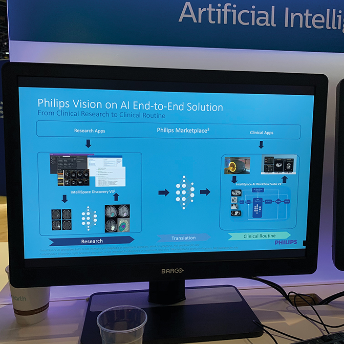
Royal Philips announced the launch of IntelliSpace AI Workflow Suite, which enables healthcare providers to seamlessly integrate AI into the imaging workflow. The AI workflow suite provides a full set of applications to integrate and centralize workflow management of AI algorithms, delivering structured results wherever they’re needed across the healthcare enterprise.
It automatically orchestrates the routing of clinical data to the appropriate AI application, analyzes the data without user interaction, then displays the results – allowing providers to capitalize on AI applications without adding complexity. IntelliSpace AI Workflow Suite is launching with more than 20 applications from both Philips and its partners.
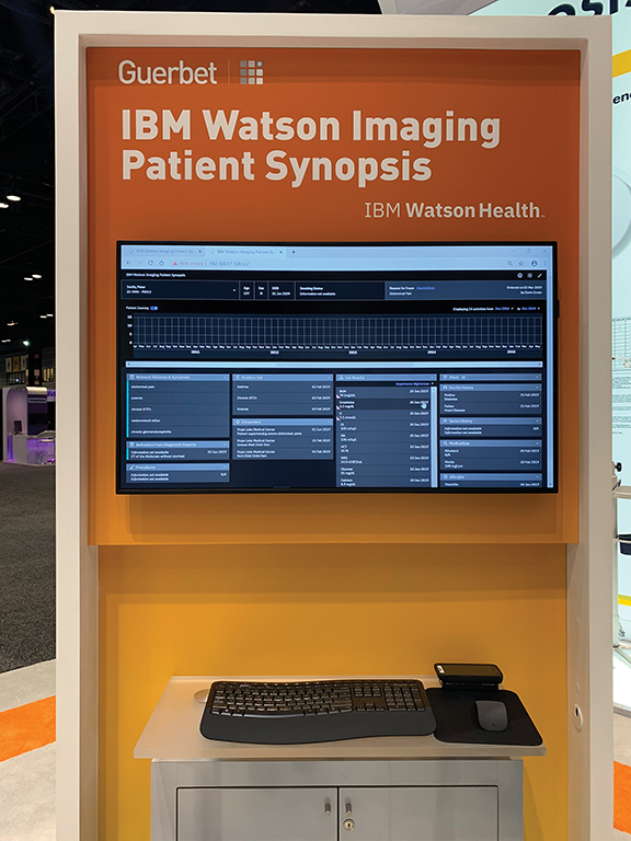
Guerbet showcased IBM Watson Imaging Patient Synopsis, a radiologist-trained AI tool that aims to extract patient information and summarize it in a tailored and specific dashboard to help inform diagnostic decisions.
“The AI engine searches for all relevant data in patients’ records and brings in data from unstructured reports to give a more holistic view of the patient,” said Leeann Essai, North American head of marketing. “Now clinicians don’t have to click through an EMR – the system surfaces relevant data so they can make a better decision quickly and efficiently.”
IBM Watson Imaging Patient Synopsis is a radiologist-trained AI tool that aims to extract relevant patient information and summarizes into a tailored and specific dashboard to help inform diagnostic decisions.
CorTechs Labs’ CEO, Chris Airriess, PhD, said the future of AI in healthcare will be powered by smart technology that can aid physicians in faster, more accurate diagnosis of neurodegenerative diseases. He called out NeuroQuant, an MR software that provides volumetric measurements of brain structures and compares them to a normative database adjusted for age, sex, and intracranial volume. It can help replace time-consuming manual processes with leading-edge automated technology that accelerates analysis so clinicians can spend more time focusing on patients.
“NeuroQuant can help doctors follow disease progression with reports tailored to diseases, such as epilepsy and multiple sclerosis,” he said. “Over time, with NeuroQuant, doctors can see how therapies for multiple sclerosis are really making a difference in their brains. It supports diagnosis through treatment follow up, which is huge.”
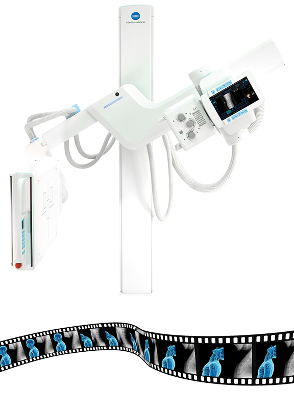
Konica Minolta Healthcare Americas, Inc., continued to highlight its focus on bringing precision medicine to primary imaging with Dynamic Digital Radiography (DDR) on the KDR Advanced U-Arm (AU) for orthopedic imaging featuring “X-ray that Moves.” Although DDR and the KDR AU have regulatory approvals, the combination of the two technologies is pending FDA clearance. When the two technologies are applied to musculoskeletal (MSK) imaging, clinicians can assess changes in the relationship of bones, ligaments, and other anatomical structures through full range of motion to evaluate the shoulders, knees, wrists, and spine.
Konica Minolta previously launched DDR for pulmonary imaging through a partnership with Shimadzu Medical Systems. In addition to producing dynamic sequences, the KDR AU also provides standard medical images for all anatomies. “DDR provides a new level of information using the common X-ray and allows the clinician to see things that they have never seen before,” says President and CEO David Widmann. “It is Smart X-ray and the foundation upon which we will continue to add advanced analytics to provide an even greater level of insight.”
KDR AU with DDR from Konica Minolta Healthcare captures a cine loop of rapidly acquired, diagnostic-quality images depicting full views of articulatory mobility.
The company also launched Rede PACS, a new PACS designed for specialty practices including orthopedic, urgent care and family practice. The Insights dashboards, initially available for Konica Minolta Healthcare’s AeroDR flat panel detectors, is now extended to provide analytics on the digital radiography CS-7 and Ultra systems and the Exa Enterprise Imaging platform. Also announced was the developed of an AI platform that will host multiple algorithms across vendors to provide a single worklist and workstation through the company’s Exa Enterprise Imaging platform.
Aidoc released its AI package for identifying and triaging stroke in CT scans will now include the CE-marked Large Vessel Occlusion AI module. The package prioritizes ischemic and hemorrhagic stroke patients in worklists by analyzing scans continuously in the background to reduce “door-to-needle” time.
Several companies announced collaborations with IBM Watson Health. Circle Cardiovascular Imaging’s cvi42, an AI-embedded single-platform solution for cardiac imaging, reading, and reporting, is now part of the IBM Imaging AI Marketplace. The platform for cardiac MR and CT, along with interventional planning and electrophysiology, provides tools to quantify and diagnose complex cardiovascular diseases. DiA Imaging Analysis Ltd announced it will introduce LVivo EF on the IBM Imaging AI Marketplace. LVivo is an AI-based quantification solution providing automated clinical data such as ejection fraction and global longitudinal strain.
MaxQ AI announced new partnerships with various original equipment manfacturers and marketplaces. Besides expanding its partnership with GE Healthcare’s PACS and Universal Viewing platforms (the solution is already available on GE’s Edison ecosystem), MaxQ’s AI ACCIPIO ICH and Stroke Platform will be integrated with Philips’ CT systems. The company also announced the availability of this solution on the Arterys Marketplace, the Nuance AI Marketplace, and the Blackford Platform.
SubtleGAD, a product of Subtle Medical, is under development for reducing gadolinium dose in MR exams. Supported by a $1.6 million National Institutes of Health Fast-Track Small Business Innovation Research (SBIR) grant, the company says the solution will use deep learning to deliver safer MRI exams without sacrificing clinical quality.
Several AI-driven cloud innovations are now available in the newest release of PowerScribe One from Nuance. Ambient Mode automatically converts free-form dictation into structured reporting, and makes possible real-time, bi-directional data synchronization with third-party platforms. A virtual assistant lets radiologists use voice commands to execute common commands, while new administrator dashboards provide real-time access to system and radiologist performance data. The company also introduced a new interface, and PowerScribe One now integrates with Nuance AI Marketplace through the solution’s image-sharing network.
For interventional fluoroscopy or cine imaging, the newly FDA-cleared FluoroShield from Omega Medical Imaging automatically collimates during a procedure to reduce field-of-view, and subsequent lower exposure to ionizing radiation. Auto ROI automatically follows device movement to minimize input requirements for the interventionalist. FluoroShield is compatible with the company’s 2020 Cardiac Flat Panel Detector.
Oxipit introduced Dynamics for its ChestEye CAD suite, which enables comparison of X-rays and provides automatically generated reports to address changes in images over time. The solution will initially support longitudinal comparison for pneumothorax, consolidation, mass, nodule, pleural effusion, pulmonary edema, and lung congestion radiological findings.
Several products under development at Care Mentor AI are designed to assist radiologists with interpretation of X-rays of the chest, foot, knee, and breast. In the breast, a neural network performs screening tasks and classifies results based on the BI-RADS algorithm. In the foot, the neural network determines the angle of the foot arch and compares it to a reference standard. Chest X-rays are screened for radiological abnormalities; recommendations are made for additional diagnostic procedures. In the knee, bone and cartilage structures, along with the width of the articular cavity, are analyzed to determine extent of osteoarthritis.
Fovia, Inc., introduced solutions that the company says will help clinicians access AI results in their existing workflows. The F.A.S.T. AI Suite provides PACS and universal viewer OEMs a way to integrate and visualize AI results within existing workflows. Also released were XStream aiCockpit, which provides access to customized, AI-driven workflows and visualizations launched from PACS, and XStream aiPlatform, a vendor-neutral ecosystem that connects AI developers, PACS and hospital systems for delivery of AI-driven tools to radiologists.
RTI-MRI+ from HealthLytix uses AI and a tissue-microstructure model to increase MR diffusion imaging visibility of restricted water in body tissue. “A key innovation of RSI-MRI+ is that it can better characterize the complexity of water diffusion in cancerous tissue, resulting in better performance,” said Nathan White, PhD, CEO of HealthLytix and coinventor of RSI. “With early-stage aggressive cancer, we need more sophisticated approaches to separate what’s important from what’s not important. In cancer, that means separating restricted diffusion from other sources of water diffusion that are less relevant.” The solution received FDA marketing clearance right before RSNA 2019.
Meanwhile, Laurel Bridge AI Workflow Suite, designed to simplify AI algorithm integration into existing clinical workflows, can manage such tasks as identifying, fetching, anonymizing, and delivering current and prior studies for use in AI algorithms or postprocessing applications. The suite can also reidentify and store AI algorithm results in an archive and provide interoperability between facilities and cloud-based AI algorithms.
TeraRecon introduced AI Sync results interchange technology for delivering interactive findings into the radiology report and downstream systems. AI Sync is an enhancement to the company’s EnvoyAI platform that prepares and presents AI results for verification before, during, or after interpretations are completed. The company also announced multiple AI-enabled workflow subscription offerings for stroke and trauma, TAVR, and cardiac MR.
Kheiron and the University of California-San Francisco (USCF) will collaborate on AI imaging research and development in UCSF’s new Center for Intelligent Imaging. Kheiron will work with UCSF’s breast imaging group to study whether the Mia breast cancer screening software can be deployed in ethnically diverse populations.
For lung CT imaging, LungPrint Discovery, from Vida, is an automated, AI-powered analysis of an inspiratory chest CT scan that flags abnormalities that may indicate emphysema, COPD, or interstitial lung disease. It includes patent-pending airway visualization for reviewing and reporting lung CT abnormalities. Designed for thin-slice, noncontrast standard chest CT scans, including those for lung screening, it is part of the company’s VIDA|vision suite of applications for lung visualization.
Volpara showcased its newly redesigned VolparaScorecard+, which provides access to three key patient risk insights: breast density assessment, an indication of suspicious findings in the mammogram, and lifetime risk of developing breast cancer. The solution incorporates Screenpoint Medical’s Transpara, an AI software that helps radiologists interpret screening mammograms. According to Volpara’s Chief Medical Officer, Monica Saini, MD, MS, the solution will “help radiologists design a personalized breast care plan for patients without disrupting their workflow.”
A new indication was cleared by the FDA for Zebra Medical Vision’s HealthCXR as part of the company’s AI1 bundle. The device can now identify and triage pleural effusions on chest X-rays.
“Based on the real-world application of this product, we saw that Zebra-Med’s automatic identification of pleural effusion on chest X-rays can play a significant role in triage,” says Dr. J.J. Visser, Radiologist and Head of Imaging IT and Value-Based Imaging at Erasmus MC University Medical Center (Netherlands). “It could be a relevant indicator for acute cardiopulmonary disease, so that clinical management can be adopted, as soon as possible, in order to provide optimal patient care.”
Citation
M B, MB M.The Ubiquity of AI at RSNA 2019. Appl Radiol. 2020; (1):32-38.
January 23, 2020