Technology and Industry: RSNA review—Part 2
Images


The technical exhibits at the 90th Scientific Assembly and Annual Meeting of the Radiological Society of North America (RSNA 2004) set 2 new RSNA records. The total number of exhibitors hit an all-time high of 690-with 134 first-time participants-and the total floor space used exceeded 455,000 square feet. The following is a brief glimpse at some of the new products showcased in Chicago, IL, November 28 through December 3, 2004.
Hologic
Hologic, Inc. (Bedford, MA) featured several women's health imaging products including a new multimodality breast imaging workstation and a high-resolution vertebral assessment tool.
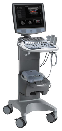
The SecurviewDX breast imaging workstation (Figure 1) is a multimodality workstation for display and interpretation of diagnostic and screening mammograms, as well as images from all other modalities, including magnetic resonance (MR), computed tomography (CT), positron emission tomography (PET), and ultrasound. The workstation is also capable of supporting computer-aided detection (CAD) programs.
"The ability to use our dedicated workstation to view digital mammograms from any vendor, as well as images for other modalities, should have a significant impact on improved workflow in breast imaging suites," said Jack W. Cumming, Chairman and CEO of Hologic. "In addition, physicians will now be able to use our package of specialized image manipulation tools on virtually any digital mammogram. These tools have been developed in close cooperation with clinical end-users to support high-quality, high-volume diagnostic case review."
The company also announced that the company's Discovery Series Densitometers (Figure 2) will now include a new modality known as Radiological Vertebral Assessment (RVA). Designed to help the physician detect vertebral fractures, RVA provides a high-resolution image of the spine as a part of the bone densitometry examination. Reimbursement for this assessment is covered under the new CPT code (76007) for vertebral fracture assessment performed using dual-energy X-ray densitometers.
Radiological vertebral assessment uses the company's proprietary OnePass fan-beam technology and radiographic standards, such as variable collimation plus single energy imaging. The company reports that the OnePass fan-beam technology uses nearly 8 times the number of detectors as competitive rectilinear dual-energy X-ray absorptiometry systems, thereby reducing imaging time by >800%.
Currently available on Hologic's Discovery series densitometers, RVA is expected to be available as an upgrade to previous generations of the company's QDR series systems in early 2005.
Barco
Also at RSNA 2004, Barco (Kortrijk, Belgium) presented upgrades to its Coronis medical display systems, personal digital assistant (PDA) support for its MediCal Administrator software, and, as a work-in-progress, a new vessel analysis software module.
The Coronis display system is now equipped with the company's 10-bit digital display controller, the BarcoMed Coronis, designed to speed workflow and enhance manipulation capabilities. The dual-head controller provides 1024 simultaneous levels of gray and features 64-bit, 66 Mhz PCI performance and image download speeds >400 Mb/sec.
Barco also added a new 2 megapixel (2MP) display with 1600 × 1200 resolution. This new display features in-plane switching (IPS) technology for enhanced viewing of angle characteristics.
In addition, Barco presented new remote calibration and problem-solving capabilities for its hospital-wide image quality management system. The new features of MediCal Administrator 2.05 include PDA support, enhanced reporting functionality, and administrator notification through the simple network management protocol (SNMP).
The new version provides system administrators with access to all medical display systems via their PDA and provides the ability to control proactive checkups from any remote location. As a result, quality assurance (QA) interventions can be performed at any time without disturbing radiology activities. It also allows Pocket PC users to view information about each display system and the QA tasks that have been performed, along with a status overview of all displays within the facility.
The company has also enhanced the program's reporting capabilities. With MediCal Administrator 2.05, QA administrators can now export all QA data reports to familiar file formats, such as Excel and PDF. The new version also includes extended notification functionality. Technicians can now automatically be notified via SNMP whenever one of the display systems performs below standard.
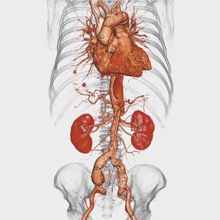
Lastly, Barco also used RSNA 2004 to preview its new vessel analysis software from its Voxar product group. The new VesselMetrix offers advanced multiplanar reconstruction (MPR) and 3-dimensional (3D) functionality for analysis of aneurysms and stenosis (Figure 3). It provides a clinical protocol-based workflow for quantitative analysis of contrast-enhanced CT angiography (CTA) and MR angiography (MRA) studies, including stenosis and stent-graft planning and surveillance.
According to the company, "the VesselMetrix module was designed to increase productivity, report key images faster, and make the clinical task of reading CTA and MRA studies easier with minimal user intervention." With this program, automated vessel finding serves as a baseline for vessel measurements and permits segmentation of vessels with 2 mouse clicks. The Live Images capabilities allow the user to prepare a study for viewing and save it for subsequent reading.
"Clinical exams, such as CTA, are now becoming a routine part of a radiologist's daily worklist, quickly replacing invasive, expensive exams such as traditional catheter angiography," said Craig Anderson, General Manager of the Voxar Product Group at Barco. "VesselMetrix provides radiologists with advanced MPR and 3D functionality to improve productivity in the visualization and analysis of selected vessels for accurate diagnosis of aneurysms and blocked arteries."
GE Healthcare
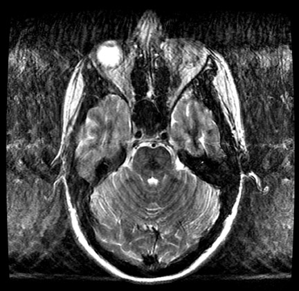
GE Healthcare (Chalfont St. Giles, UK) used RSNA 2004 to launch its first high-definition magnetic resonance (HDMR) system. According to the company, this system was designed to improve speed and image quality for patients with uncontrollable movement, such as those with Parkinson's disease and children who do not respond to sedation (Figure 4).
This new technology, available on the Signa 1.5T and 3.0T MR systems, enables massively simultaneous imaging in multiple channels in increments of 16. Image-processing power increases in proportion as sets of 16 channels are added.
GE's proprietary EXCITE technology previously enabled 3 high-resolution, targeted MR applications: VIBRANT for bilateral breast imaging in a single examination; TRICKS for MRA of the legs; and PROPELLER for high-quality, motion-resistant brain imaging.
"Now with HD, we've an extremely fast data processing engine coupled with high-density surface coils and extremely accurate gradients," said Dennis Cooke, GE Healthcare's Vice President, Global MR Business. "The result is a unique balanced acquisition architecture, with individual receive channels connected to dedicated reconstruction engines. This allows us to develop entirely new applications that are available only with HDMR." The new targeted studies include high-resolution imaging of the liver with greater coverage and shorter breath-holds, MR echo real-time imaging of the heart without the need for breath-holding or ECG gating, and enhanced definition of peripheral vascular imaging of the lower leg and foot with a new 32-element peripheral vascular coil.
"Dedicated high-density coils facilitate throughput and enable high-definition scanning, resulting in dramatic images reminiscent of those provided by high-definition television," added Lawrence N. Tanenbaum, MD, Section Chief MRI, CT, and Neuroradiology, New Jersey Neuroscience Institute (Edison, NJ) and a member of the editorial board of this journal.
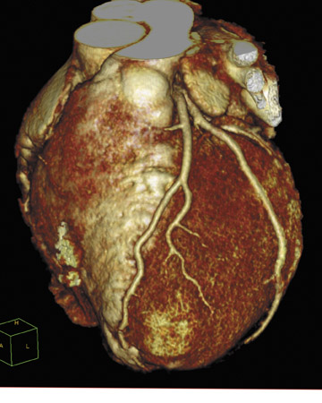
The company also introduced its next-generation volume CT (VCT) scanner, the LightSpeed VCT. This new system, with a 64-channel detector, 0.35-sec rotation time, and 40-mm coverage, allows for scanning of the heart in 5 beats.
The first LightSpeed VCT was installed at Froedtert Hospital, Milwaukee, WI, in 2004. "It is somewhat amazing that an accurate 3D image of the coronary arteries and sites of focal disease can be obtained from a beating heart," said W. Dennis Foley, MD, Chief of Digital Imaging at Froedtert and Professor of Radiology, the Medical College of Wisconsin, Milwaukee. Foley noted, "The LightSpeed VCT system does this using rapid high-resolution imaging gated to the patient's heart cycle."
According to the company, the LightSpeed VCT can attain 43-msec temporal resolution, effectively freezing the motion of the heart. In a single rotation, the system creates 64 submillimeter images, totaling 40 mm of anatomic coverage, which are combined to form a 3D view of the patient's anatomy. The system is also able to scan the entire body in 10 seconds.
The 5-Beat Cardiac examination application for this system allows the user to obtain high-quality images of coronary arteries at submillimeter resolution in only 5 beats of the heart for rapid diagnostic evaluation of arterial stenosis. The Triple RuleOut application was designed for use in patients who present to the emergency department with acute chest pain. Such patients can undergo a single scan with the LightSpeed VCT to search for evidence of heart attack, pulmonary embolism, or aortic dissection. For stroke patients, the LightSpeed VCT offers rapid perfusion studies of the brain, enabling doctors to make a quick diagnosis of stroke and determine the extent of damage.
3TP Imaging Sciences
3TP Imaging Sciences (White Plains, NY) introduced its new postprocessing computer analysis software for dynamic contrast-enhanced MR images of the prostate at RSNA 2004.
This proprietary technology, developed by Professor Hadassa Degani, Head of Magnetic Resonance for Biomedical Research at the Weizmann Institute, Rehovot, Israel, was designed for use in the preoperative staging of biopsy-proven prostate cancer. The product includes several new features, including a major upgrade of the motion-correction software, which processes data in one third of the time previously required.
The software is an extension of the breast imaging software package introduced by 3TP and is based on the wash-in and wash-out properties of contrast media in tissue. The 3TP formula measures tissue permeability and extracellular volume fraction on a pixel-by-pixel basis at 3 distinct time points. The software assigns a color to each pixel, which enables the radiologist to rapidly identify suspicious areas for further analysis (Figure 5).
"3TP software provides a valuable tool to establish an accurate diagnosis of prostate staging, and I am tremendously excited about the potential to use 3TP to improve brachytherapy planning and to calculate tumor volume measurements," said B. Nicolas Bloch, MD, Research Fellow in Radiology, Harvard Medical School, Boston, MA, and Instructor in Radiology, Medical University of Vienna, Austria.
Suros Surgical Systems
Suros Surgical System, Inc. (Indianapolis, IN) introduced 4 new breast biopsy devices at RSNA 2004, including an all-in-one vacuum-assist-ed system, the ATEC Sapphire, which is compatible with all primary diagnostic imaging modalities, including MR. The company also unveiled the ATEC Pearl, the ATEC MRI Access petite needle, and the ATEC TriMark marker deployment system.
The ATEC Sapphire provides vacuum-assisted minimally invasive tissue excision when used with ultrasound, stereotactic guidance, or MRI (Figure 6). The lower-priced ATEC Pearl is similar but does not include the MR compatibility. Both systems are 25% smaller than the previous versions and are capable of obtaining a core nearly every 4.5 seconds, or 14 cores per minute. The completely closed systems are compatible with both 9- and 12-g needle options and are operated via the use of a single foot pedal that initiates the start and stop of the 7ounce handpiece.
The ATEC MRI Access 9-g petite needle has a unique hemispherically shaped tip that allows physicians to access biopsy targets on breasts compressed to as little as 22 mm without concern for skin perforation. According to the company, this gives women with small or "thin" breasts the option to choose a needle biopsy over an open surgical procedure. The petite needle is compatible with both the ATEC Sapphire and the ATEC Pearl.
The ATEC TriMark is a marker and deployment system that offers permanent visualization in all 3 primary breast imaging modalities. It is composed of implant-grade, biocompatible titanium and is available in 2 lengths. The TriMark 13 is used for ultrasound and MR-guided biopsies, while the TriMark 36 is for use in stereotactic-guided procedures. The marker is sized to fit both the 9- and the 12-g needles.
Citation
. Technology and Industry: RSNA review—Part 2. Appl Radiol.
February 10, 2005