Study Shows 90% Diagnostic Accuracy of Lung Nodules with Philips Technology
 Radboud university medical center (Radboudumc) has set a new milestone in the diagnostic accuracy of navigation bronchoscopy using Royal Philips’ Lung Suite combined with Azurion, the company’s Image Guided Therapy System.
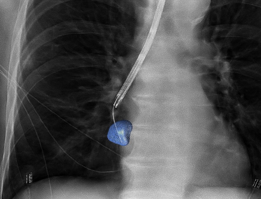
A clinical study conducted by Radboudumc reported the diagnostic accuracy and procedural radiation dose for patients undergoing an endobronchial lung biopsy supported by Philips Lung Suite, a solution that uses 3D imaging with augmented fluoroscopy to support high precision diagnosis and minimally-invasive therapy in one room. Diagnostic accuracy of 90% was reported while reducing the average total effective radiation dose per procedure by more than half from 47.5 Gy·cm2 (effective dose: 14.3 mSv) to 25.4 Gy·cm2 (effective dose: 5.8 mSv). The median long-axis diameter of the 248 lesions navigated to during the study was 13 mm (range 5-65mm). The results of the study were published in the October 2021 issue of the Journal of Bronchology & Interventional Pulmonology.
“The airways and lungs are very challenging places to biopsy, so we need good navigation to make sure we reach the target location,” said Dr. Erik van der Heijden, Pulmonologist and Associate Professor of Interventional Pulmonary Diseases at Radboudumc, who led the study. “Philips’ 3D imaging solution allows us to create real-time 3D visualizations enabling us to follow the correct path from different angles to where we need to be, which is particularly useful for very small abnormalities. Our study confirmed that it increases the accuracy and safety of the biopsy procedure, improving results for patients. Philips’ technology also offers the promising outlook that we could not only diagnose but also immediately treat these early-stage patients using novel procedures such as tumor ablation.”
The research being carried out at Radboudumc focuses on the diagnosis of early-stage lung cancer lesions, typically detected during routine screening of high-risk patient groups or discovered and reported as so-called “incidental findings” when a patient has a chest scan for some other reason. A tissue sample typically then needs to be obtained that a pathologist can use to make a definitive diagnosis.
A new method is image-guided endobronchial lung biopsy, during which a clinician uses live image guidance to maneuver through the airways in the lungs to advance a catheter towards the lesion. However, the airways in the lungs are a complex network that requires accurate navigation to remove the required tissue sample. Until now, image-guided endobronchial lung biopsy has only really been effective when an abnormality or tumor is located in or near one of the larger airways in the lungs, and even then it is challenging for clinicians to accurately navigate when looking at a 2D grayscale fluoroscopy image while the lungs are moving as the patient breathes.
Philips Lung Suite enables all-in-one lung cancer diagnosis and treatment. It provides advanced real-time 3D imaging with augmented fluoroscopy on the company’s Image Guided Therapy System – Azurion, combined with dedicated software. With Philips’ Cone Beam CT imaging, the X-ray detector rotates around the patient to generate a CT-like image in around five seconds, providing clinicians with a high-resolution 3D view of the target lesion and other anatomical structures. This allows the clinician performing the biopsy procedure to be continually guided by high-quality real-time imaging to advance a catheter towards the lesion through a bronchoscope. Once done, its position can be confirmed in real-time using the same imaging modality and a biopsy sample removed.
Related Articles
Citation
Study Shows 90% Diagnostic Accuracy of Lung Nodules with Philips Technology. Appl Radiol.
October 7, 2021