Study Reports Dynamic Digital Radiography Quantitatively Diagnoses Diaphragm Dysfunction
Images
A case study by clinicians in the pulmonary and radiology departments at ASST Fatebenefratelli Sacco (Milan, Italy) demonstrates the use of Dynamic Digital Radiography (DDR) to help definitively diagnose diaphragm dysfunction. The study, published in the journal Diagnostics, highlights the clinical value of DDR through its unique ability to evaluate diaphragm movement in real time and integrate dynamic functional information with static anatomical data to provide a quantitative assessment of diaphragmatic movement, including excursion and speed.
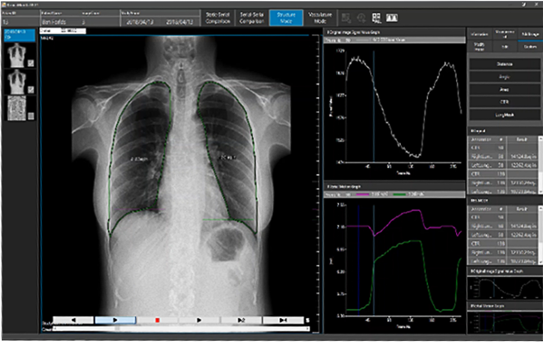
DDR was used to evaluate a 46-year-old male with a past history of smoking and a 2021 SARS-CoV-2 infection for unexplained dyspnea and reduced exercise tolerance. Spirometry values were below predicted values and a standard chest radiograph depicted an elevated right hemidiaphragm that was not present on a prior CT examination during his SARS-CoV-2 infection.
With the dynamic functional imaging capability of DDR, the authors could visualize thoracic and pulmonary motion and track diaphragm movement. Additional post-processing of the DDR data using Konica Minolta’s Intelligent Workstation (IWS, also known as DI-X1) enabled the measurement of diaphragmatic motion and overall lung mobility, highlighting regional differences in ventilation of both lungs. As a result of the DDR examination, the patient was referred to a thoracic surgeon for possible plication of the right diaphragm.
“Diagnosing diaphragm dysfunction is challenging due to its varied symptoms and causes. While conventional chest X-rays and ultrasound can offer clues, additional imaging and tests are often needed for a precise diagnosis,” says Michaela Cellina, Head of Imaging Research and a radiologist with ASST Fatebenefratelli Sacco. “Dynamic Digital Radiography is an innovative imaging technique that can be captured at the same time as a traditional static chest X-ray, providing valuable real-time insights for a quick and confident diagnosis.”
DDR is the only FDA-cleared radiography solution that enables visualization of anatomy in motion. It acquires up to 15 sequential radiographs per second and processes them as a cine loop, enabling clinicians to observe the physiological cycle as well as individual radiographs. No physician presence is required, radiation dose is lower than an average fluoroscopy exam and the dynamic images can be captured with the patient sitting, standing or on a table.
“Konica Minolta congratulates Dr Michaela Cellina, Dr Elisa Calabro and their colleagues on the publication of this case study that demonstrates the ability of one examination – DDR – to deliver a definitive diagnosis of the cause of dyspnea,” says John Sabol, PhD, Clinical Research Manager, Konica Minolta Healthcare. “Adding DDR to a conventional chest X-ray, which is routinely acquired in these cases, may eliminate the need for additional tests or imaging studies, helping reduce costs to the patient and health system, and enable a more rapid diagnosis.”
Related Articles
Citation
Study Reports Dynamic Digital Radiography Quantitatively Diagnoses Diaphragm Dysfunction. Appl Radiol.
February 14, 2025