Standard MRI protocols diagnose pancreas divisum as well as 3D-MRCP, in less time, with fewer artifacts
Three-dimensional magnetic resonance cholangiopancreatography (3D-MRCP) is the examination most frequently ordered to diagnose pancreas divisum, a congenital anomaly in the ducts of the pancreas that occurs in up to 14% of the population. However, 3D-MRCP has lengthy acquisition times and is prone to motion artifacts.
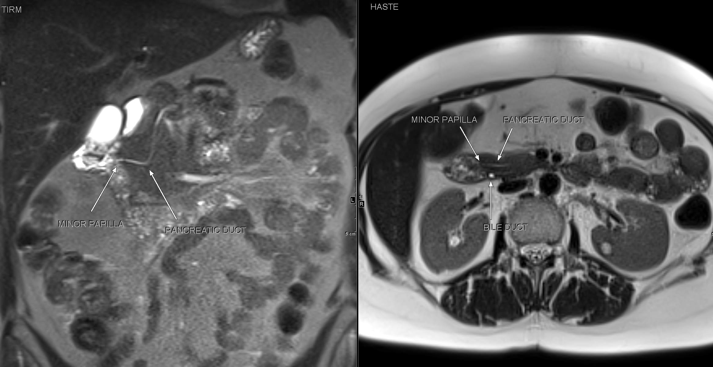
Example of pancreas divisum diagnosis using routine non-mRCP T2-weighted sequences (TIRM, HASTE).
T2-weighted MR imaging sequences, on the other hand, are diagnostically “non-inferior” to 3D-MRCP, and may produce fewer artifacts, according to researchers at Heidelberg University Hospital in Germany.
In pancreas divisum, the dorsal and ventral ducts fail to fuse into a single duct, and digestive juices drain via the dorsal duct and the narrow minor papilla instead of through the ventral duct and via the major papilla. While most individuals with the condition are asymptomatic, a small percentage may experience abdominal pain, nausea, and acute or chronic pancreatitis.
Hypothesizing that non-MRCP, T2-weighted sequences frequently included in routine abdominal MRI protocols could produce high-quality diagnostic images to effectively diagnose pancreas divisum, the researchers reviewed 342 consecutive patients who had abdominal MRI scans that included 3D-MRCP over a 10-month period and who did not have a history of pancreatobiliary surgery.
The cases included the images of 172 men and 170 women, aged 35-67 years. Clinical indications for imaging included intrapancreatic inflammatory disease, intrapancreatic tumor, extrapancreatic non-oncological disease, and extrapancreatic tumor. Ten percent were diagnosed with pancreas divisum.
In April 19, 2019, online edition of BMC Medical Imaging, the authors explained that the 3D- MRCP was a coronal, respiration-navigated T2-weighted sequence with 1.5-mm slices. The non-MRCP sequences, meanwhile, included a coronal inversion recovery sequence (TIRM) with 6-mm slices and a transverse single shot turbo spin echo sequence (HASTE) with 4-mm slices.
One radiologist assessed the three sequences for the presence of pancreas divisum. Two radiologists subsequently reviewed these in consensus while also assessing image quality and rating the presence of motion artifacts on the 4-point Likert scale.
Led by Tim Frederik Weber, MD, of the department of diagnostic and interventional radiology, the team reported that each of the two non-MRCP T2-weighted sequences had greater sensitivity (92.5% for TIRM and 98.1% for HASTE) and specificity (93.9% for TIRM and 97% for HASTE) than the 3D-MRCP sequences (81.2% and 69.7%, respectively). The negative predictive value for the combined non-MRCP sequences was 99.9%.
“Both TIRM alone and HASTE alone as well as the combination of TIRM and HASTE were non-inferior to 3D-MRCP for diagnosis of pancreas divisum,” they wrote.
Each non-MRCP sequence provided images with higher evaluability with respect to motion artifacts and sufficiency of duct depiction. Only 2.3% of the HASTE-acquired images and 7.3% of the TIRM-acquired images were not evaluable, compared to 20.2% of the images acquired by 3D-MRCP. The authors attributed the difference to some patients’ limited capability for shallow constant breathing, even when 3D-MRCP acquisition was synchronized to respiration.
“If 3D-MRCP is not evaluable, non-MRCP T2-weighted sequences frequently included in routine abdominal MRI protocols are sufficient to illustrate pancreatic duct anatomy in regards of pancreas divisum,” said Dr. Weber. He told Applied Radiology that Heidelberg’s radiology department still performs 3D-MRCPs, but relies on TIRM and HASTE for pancreas divisum diagnosis or exclusion, especially where 3D-MRCP image quality is insufficient.
REFERENCE
- Bogveradze N, Hasse F, Mayer P, et al. Is MRCP necessary to diagnose pancreas divisum? BMC Med Imaging. Published online April 19, 2019. 19(1):33. doi: 10.1186/s12880-019-0329-1.