SNMMI ’21: Image of the Year Goes to PET Imaging Measuring Cognitive Impairment in COVID-19 Patients
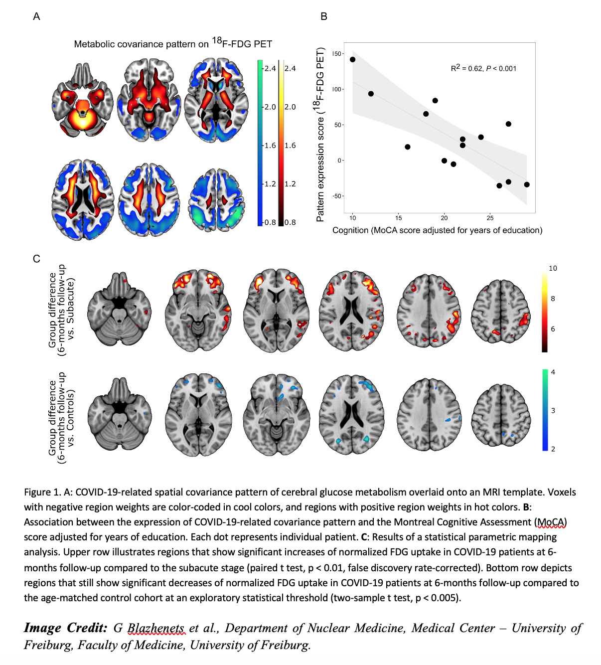 The Society of Nuclear Medicine and Molecular Imaging (SNMMI) 2021 Henry N. Wagner, Jr., Image of the Year was awarded to an image that accurately measured the effects of COVID-19 on the brain with PET. In the study, newly diagnosed COVID-19 patients, who required inpatient treatment and underwent PET brain scans, were found to have deficits in neuronal function and accompanying cognitive impairment, and in some, this impairment continued six months after their diagnosis. The selected image (pictured) detailed depiction of areas of cognitive impairment, neurological symptoms and comparison of impairment over a six-month time frame.
Each year, SNMMI chooses an image that best exemplifies the most promising advances in the field of nuclear medicine and molecular imaging. The state-of-the-art technologies captured in these images demonstrate the capacity to improve patient care by detecting disease, aiding diagnosis, improving clinical confidence, and providing a means of selecting appropriate treatments. This year, the SNMMI Henry N. Wagner, Jr., Image of the Year was chosen from more than 1,280 abstracts submitted to the meeting and voted on by reviewers and the society leadership.
“As the SARS-CoV-2 pandemic proceeds, it has become increasingly clear that neurocognitive long-term consequences occur not only in severe COVID-19 cases, but in mild and moderate cases as well. Neurocognitive deficits like impaired memory, disturbed concentration and cognitive problems may persist well beyond the acute phase of the disease,” said Ganna Blazhenets, PhD, a post-doctoral researcher in Medical Imaging at the University Medical Center Freiburg, in Freiburg, Germany.
To study cognitive impairment associated with COVID-19, researchers carried out a prospective study on recently diagnosed COVID-19 patients who required inpatient treatment for non-neurological complaints. A cognitive assessment was performed, followed by imaging with 18F-FDG PET if at least two new neurological symptoms were present. By comparing COVID-19 patients to controls, the Freiburg group established a COVID-19-related covariance pattern of brain metabolism with most prominent decreases in cortical regions. Across patients, the expression of this pattern showed a very high correlation with the patients’ cognitive performance.
Follow-up PET imaging was performed six months after the initial COVID-19 diagnosis.
Imaging results showed a significant improvement in the neurocognitive deficits in most patients, accompanied by an almost complete normalization of the brain metabolism.
“We can clearly state that a significant recovery of regional neuronal function and cognition occurs for most COVID-19 patients based on the results of this study. However, it is important to recognize the evidence of longer-lasting deficits in neuronal function and accompanying cognitive deficits is still measurable in some patients six months after manifestation of disease,” noted Blazhenets. “As a result, post-COVID-19 patients with persistent cognitive complaints should be presented to a neurologist and possibly allocated to cognitive rehabilitation programs.”
“18F-FDG PET is an established biomarker of neuronal function and neuronal injury,” stated SNMMI’s Scientific Program Committee chair, Umar Mahmood, MD, PhD. “As shown the Image of the Year, it can be applied to unravel neuronal correlates of the cognitive decline in patients after COVID-19. Since 18F-FDG PET is widely available, it may therefore aid in the diagnostic work-up and follow-up in patients with persistent cognitive impairment after COVID-19.”
Related Articles
Citation
SNMMI ’21: Image of the Year Goes to PET Imaging Measuring Cognitive Impairment in COVID-19 Patients. Appl Radiol.
June 15, 2021