Retroperitoneal Ancient Schwannoma
Images


Case Summary
A middle-aged woman presented with vaginal bleeding. The physical exam was unremarkable, but ultrasound revealed two cysts measuring up to 3.2 cm on the patient’s right ovary. The patient underwent diagnostic laparoscopy. Intraoperatively, the ovaries appeared normal; however, a right retroperitoneal mass was noted.
Imaging Findings
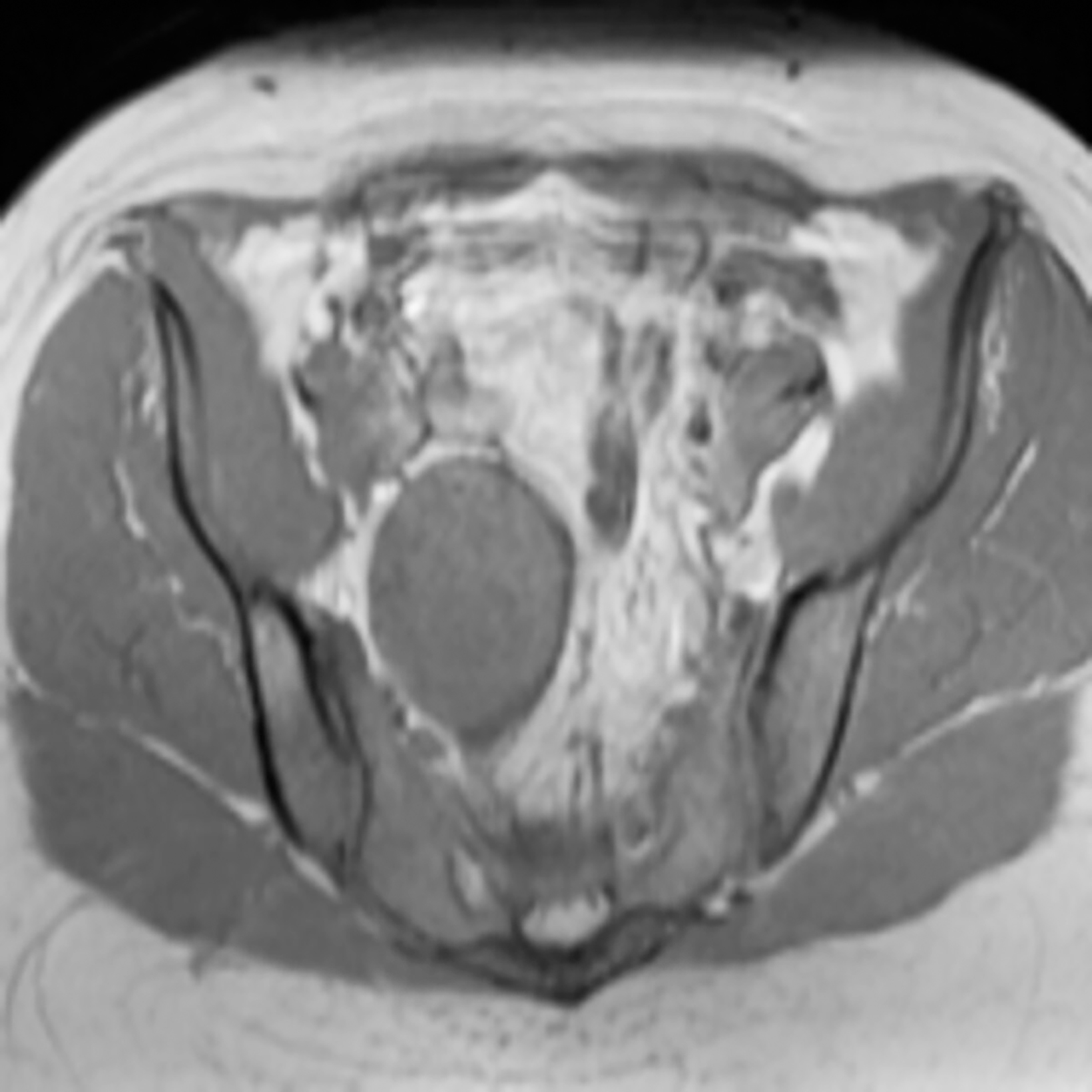
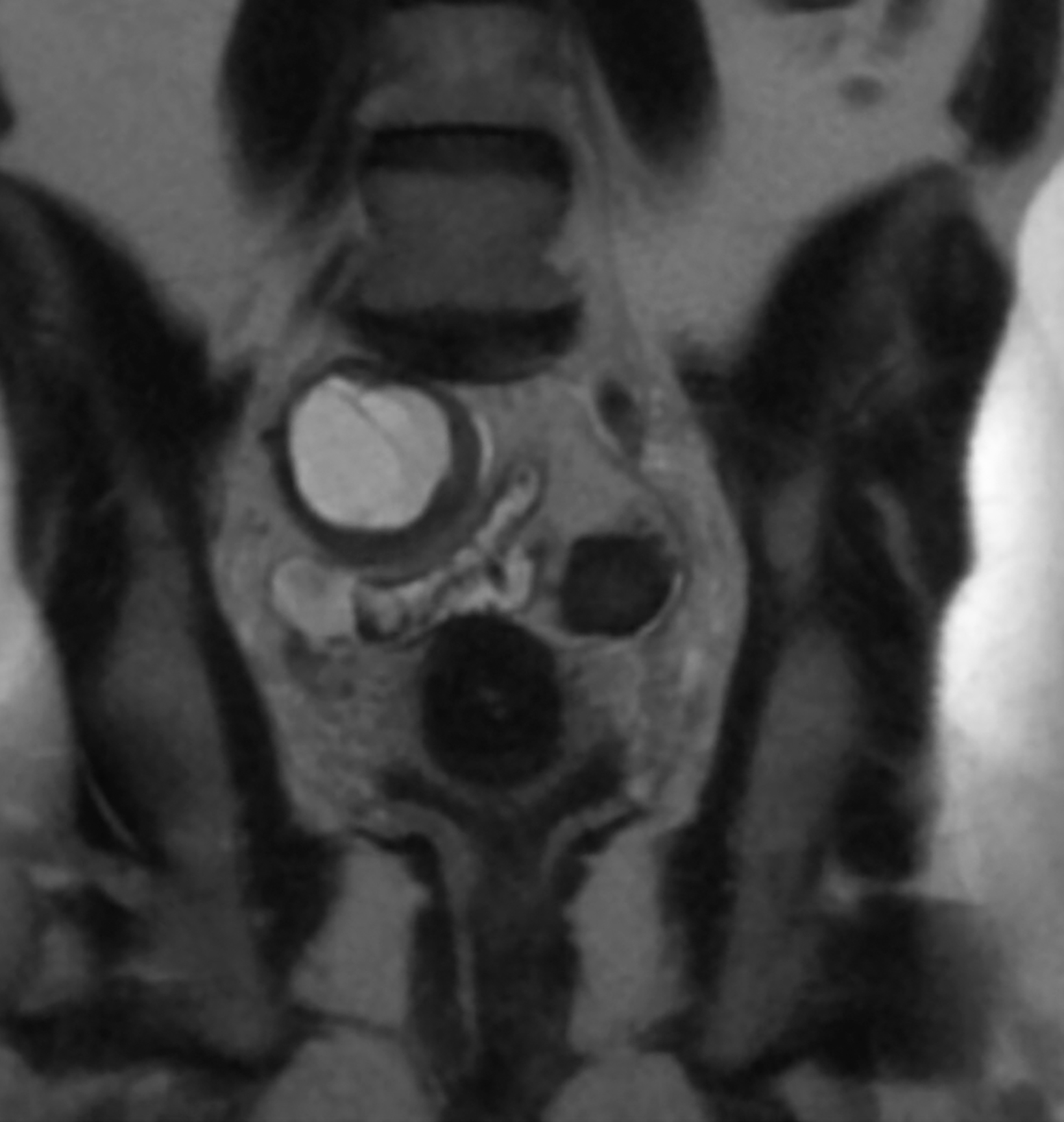
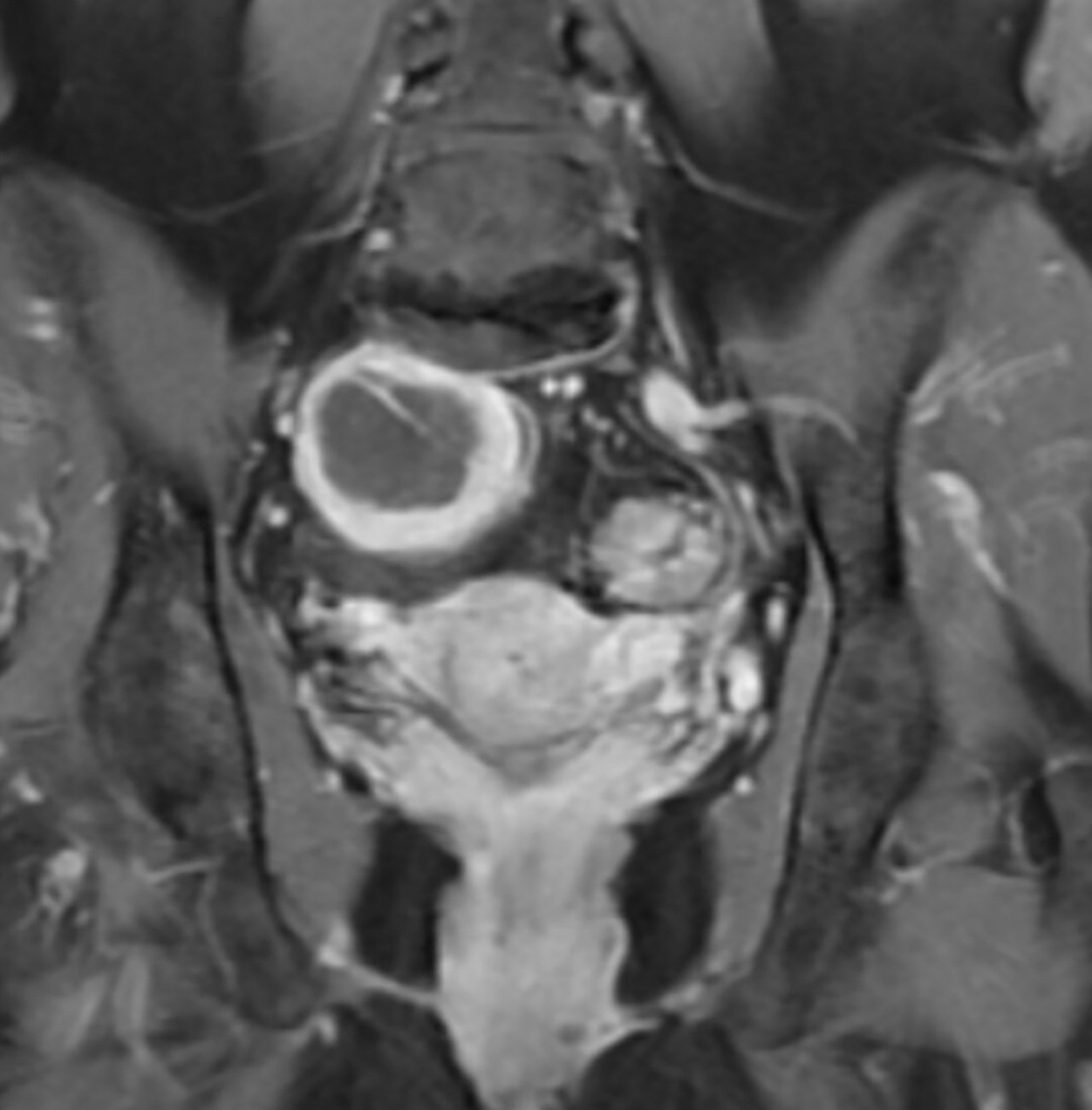
Magnetic resonance imaging (MRI) of the pelvis revealed a 5cm right retroperitoneal cystic mass. The mass was primarily isointense to muscle on T1 images (Figure 1) and hyperintense on T2 images (Figure 2). Thick-walled rim enhancement followed gadolinium administration (Figure 3). There was no diffusion restriction.
A 5.4 × 5.3 × 5.0 cm capsulated cystic mass was excised via an open transperitoneal technique. Histologic examination revealed a poorly cellular, bland neoplasm in variably dense fibrotic stroma. The cells were narrow and elongated with tapered ends interspersed with collagen fibers. Mitotic figures were not identified in the mass. Immunohistochemical staining was strongly and diffusely positive (3+, 100%) for S100, and negative for CD 34 and desmin.
Diagnosis
Retroperitoneal ancient schwannoma
Discussion
Among the most common soft-tissue tumors, schwannomas are benign tumors that arise from the Schwann cells of the nerve sheath and present with symptoms of pain or paresthesia. Ancient schwannoma, a degenerative neurilemmoma, is a subtype characterized by degeneration and diffuse hypocellular areas that are believed to result from the long time required for schwannomas to develop. 1
Only 0.75% to 2.6% of ancient schwannomas occur in the retroperitoneum. 2 They generally become very large before producing symptoms owing to mass effects. 3
Retroperitoneal schwannomas are usually solid, encapsulated tumors that originate in the paravertebral region and are more likely to undergo spontaneous degeneration and hemorrhage compared to schwannomas arising in the head, neck, and extremities. 2
These cases are typically diagnosed in patients between 40 and 60 years of age, with a male-female ratio of 2:3. 2 Diagnosing retroperitoneal schwannomas is difficult. One study reported that symptoms were nonspecific, and neurologic symptoms were rare. 4 Symptoms include vague abdominal pain, flank pain, hematuria, headache, secondary hypertension, and recurrent renal colic pain. 5 Preoperative diagnosis is difficult because of the absence of pathognomonic features. 4
Ultrasonography is a useful and inexpensive modality for detecting these tumors. Computed tomography (CT) scans can reveal well-defined low or mixed attenuation with cystic necrotic central areas. Cystic changes are seen more commonly in retroperitoneal schwannomas than in other retroperitoneal tumors. 5 MRI can be used to better characterize large retroperitoneal tumors because of the superior visualize the origin, vascular architecture, and involvement of other organs. 2 CT-guided biopsy may be helpful when samples contain enough Schwann cells for microscopic visualization. Many investigators do not recommend preoperative biopsy because of its risks for hemorrhage, infection, and tumor seeding. 6
Macroscopically, schwannomas are solitary, well-circumscribed, firm, and smooth-surfaced tumors. 1 Histologically, they are composed of Schwann cells with regions of high and low cellularity termed Antoni A and Antoni B areas, with a diffuse positivity of S100 protein. 4 The presence of degenerative changes, such as cyst formation, hemorrhage, calcification, and hyalinization, classifies these tumors as ancient schwannomas. Microscopically, Antoni A and B areas, and S100 positivity with cyst formation were seen in our case.
The differential diagnosis of retroperitoneal schwannomas includes neurofibroma, paraganglioma, pheochromocytoma, liposarcoma, malignant fibrous histiocytoma, lymphangioma, and hematoma. 4 Cystic degeneration, however, is the strongest indicator for ancient schwannoma.
In otherwise healthy patients, complete excision is the preferred treatment. 1 Malignant transformation is extremely rare, and recurrences are uncommon following surgical resection, although postsurgical monitoring is recommended. 2
Conclusion
Ancient schwannomas are degenerative peripheral nerve sheath tumors that rarely occur in the retroperitoneum. A diagnosis of ancient schwannoma should be entertained when there is a heterogeneous, well-encapsulated mass in the retroperitoneum.
References
- Isobe K, Shimizu T, Akahane T, Kato H. Imaging of ancient schwannoma. AJR Am J Roentgenol. 2004;183(2):331–336.
- Çalişkan S, Gümrükçü G, Kaya C. Retroperitoneal ancient schwannoma: a case report. Rev Urol. 2015;17(3):190–193.
- Choudry HA, Nikfarjam M, Liang JJ, et al. Diagnosis and management of retroperitoneal ancient schwannomas. World J Surg Oncol. 2009;7:12.
- Goh B, Tan YM, Chung YF, et al. Retroperitoneal schwannoma. AJ Surg. 2006;192:14–18.
- Cury J, Coelho RF, Srougi M. Retroperitoneal schwannoma: case series and literature review. Clinics. 2007; 62:359–362.
- Daneshmand S, Youssefzadeh D, Chamine K, et al. Benign retroperitoneal schwannoma: a case series and review of the literature. Urology. 2003;62:993-997.
References
Citation
N N, EI A.Retroperitoneal Ancient Schwannoma. Appl Radiol. 2021; (6):45-47.
November 6, 2021