Radiology Working Group Publishes Recommendations for Idiopathic Pulmonary Fibrosis
 The Radiology Working Group of the Pulmonary Fibrosis Foundation has published recommendations for image interpretation in patients with suspected idiopathic pulmonary fibrosis (IPF). Appearing online February 25, 2021 in Radiology: Cardiothoracic Imaging, the authors describe critical imaging patterns of IPF – including confounding finding with other fibrotic lung disease – pitfalls in imaging classifications and reporting standards.
Based in Chicago, the Pulmonary Fibrosis Foundation is a nonprofit organization founded in 2000 dedicated to identifying effective PF treatments and assisting those living with the disease. The Pulmonary Fibrosis Foundation developed a network of care centers nationwide to promote early diagnosis of pulmonary fibrosis. The organization has also created a patient registry and biorepository to assist researchers, clinicians and scientific leaders to serve as a resource and provide data for improving evidence-based care guidelines.
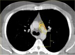
In the recommendations, the authors review imaging findings and patters associated with IPF and other fibrotic lung diseases. Although chest radiography is often utilized as an initial screening test, thin-slice CT is the primary diagnostic tool for categorization and continued follow-up of patients with IPF. The authors provide key patterns and definition for patients with and without IPF. CT images are available to download for further analysis by the reader.
According to the Pulmonary Fibrosis Foundation, pulmonary fibrosis is a classification of more than 200 different lung diseases with a similar appearance. IPF is a scarring of the lung due to unknown causes. The scarring pattern is typically referred to as usual interstitial pneumonia (UIP). The new recommendations also provide guidance on the interpretation images as the UIP pattern can be found in other health conditions including chronic HP, drug toxicity, asbestosis, and CTDs such as rheumatoid arthritis.
Related Articles
Citation
Radiology Working Group Publishes Recommendations for Idiopathic Pulmonary Fibrosis. Appl Radiol.
March 1, 2021