Prostatic utricle cyst with chronic prostatitis
Images

CASE SUMMARY
A 48-year-old army officer was referred to the radiology department for MRI of prostate to rule the cause of post void dribbling. There was no history of trauma or infection in the past. The patient was otherwise healthy and there was no other associated significant medical history. Digital rectal examination revealed a large firm non-tender midline swelling in the prostate. Lab investigations were within normal limits.
IMAGING FINDINGS
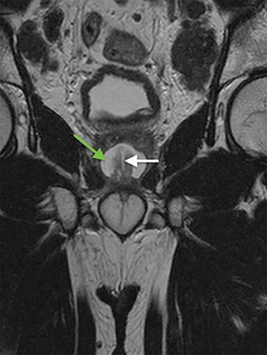
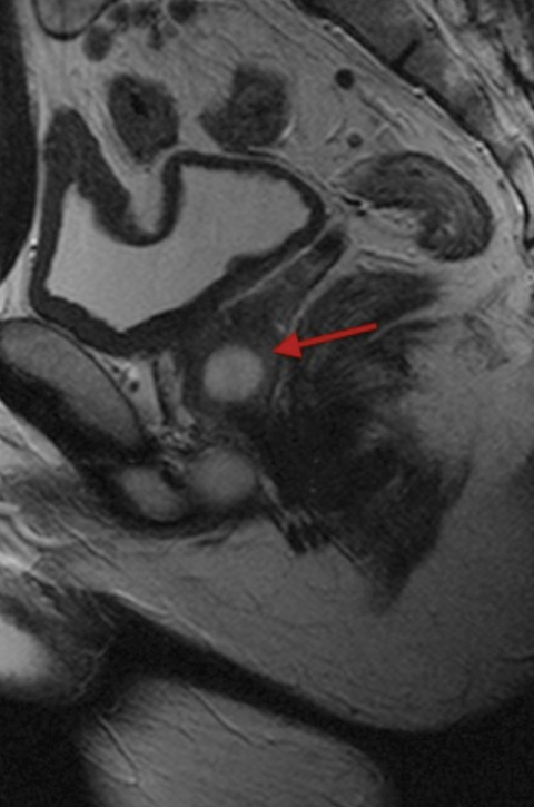
Prostate MRI was performed on 1.5T Siemens Symphony MR scanner for the diagnosis and better characterization of the pathology. A well-defined oval cystic lesion measuring 2 × 2.5 × 1.8 cm was noted within the prostate located in the midline, which was communicating with the prostatic urethra (Figures 1a, 1b). The peripheral zone of the prostate was thinned out and appeared diffusely hypointense on T2WI, suggestive of chronic prostatitis (Figure 2).
DIAGNOSIS
Prostatic utricle cyst with chronic prostatitis
DISCUSSION
Prostatic utricle is a remnant of Mullerian duct in males.1 Utricle is derived from the Latin word “pouch,” which forms a cul-de-sac 6 mm long, located in the verumontanum between the two ejaculatory ducts that projects upward and backward into the substance of the prostate. Prostatic utricle cyst results from focal dilatation of the prostatic utricle ranging from few millimeters to 2 cm.1 The incidence of prostatic utricle cyst is 11% to 14% in association with hypospadias or intersex anomalies and up to 50% in the presence of perineal hypospadias.2 Prostatic utricle cyst is usually seen during the first to second decades of life, with a mean age range of 26 years. Very few cases have been reported in the literature.
Most prostatic utricular cysts are asymptomatic, especially when small. When large, symptoms typically consist of urinary incontinence, recurrent infections, or stone formation. Malignant degeneration has been reported in 3% of prostatic utricles with a peak incidence in the fourth decade of life.3
The diagnosis is essentially based on imaging. On VCUG and RGU, prostatic utricle cyst is diagnosed when spontaneous opacification of a diverticulum arising from the prostatic urethra is seen.4 On ultrasonography, a well-defined midline cystic lesion is seen posterior to the urinary bladder. MRI depicts the utricle as a cystic lesion in the substance of the prostate gland posterior to the urinary bladder with a typical midline location. Communication of the prostatic midline cyst with the prostatic urethra is typically noted.1 Surgical resection is the treatment of choice.
The differential diagnosis to be considered on imaging appearance include bladder diverticulum, urachal cyst, Mullerian duct cyst, or a Seminal vesicle cyst. Prostatic utricle cyst communicates with the prostatic urethra and are located in the midline, posterior to the bladder, and anterior to the rectum, as described in this case. Mullerian duct cysts, on the other hand, do not typically communicate with the prostatic urethra, although they are also midline. Urachal cysts are clearly distinguished from a prostatic utricle cyst due to their anterior relationship to the urinary bladder while seminal vesicle cysts are located lateral to midline and are usually unilateral.1
CONCLUSION
Irrespective of the age of presentation, prostatic utricle cyst should be ruled out in a male patient with clinical history of urinary incon tinence.
REFERENCES
- Curran S, Akin O, Agildere AM, Zhang J, Hricak H, Rademaker J. Endorectal MRI of prostrate and periprostratic cystic lesions and its mimics. AJR. 2007;188:1373-1379.
- Mukha R, Sriram K, Ganesh G. Prostatic utricle cyst – a case report and review of current literature. The Internet J Urol. 2009;6.
- Johnson D, Parikh K, Schey W, Mar W. MRI in diagnosis of a giant prostatic utricle. Case Reports in Radiology. 2014;1-3.
- Berrocal T, Lopez-Pereira P, Arjonilla A, Gutierrez J. Anomalies of the distal ureter, bladder, and urethra in children: embryologic, radiologic, and pathologic features. Radiographics. 2002;22:1139-1164.
Citation
K C, S M, UC G.Prostatic utricle cyst with chronic prostatitis. Appl Radiol. 2016; (5):46-47.
May 1, 2016