MRI of 75-year-old female with confusion, weakness, speech problems, and visual deficit
Images
Case Summary
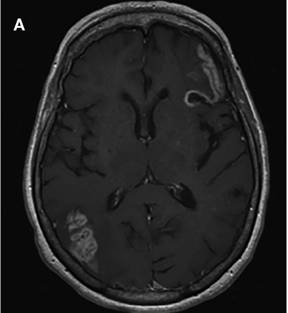
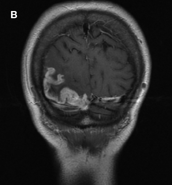
A 75-year-old woman presented with sudden onset con- fusion, weakness, expressive speech problems, and visual deficit following cardiac catheterization 3 weeks earlier. She also had a history of breast cancer with radiation therapy. An MRI of the head was performed on a 3T GE scanner. The patient received 14 mL of ProHance® (gadoteridol) administered intravenously without complication. Post-enhanced axial and coronal T1-weighted images are shown.
Imaging Findings
There is thick, curvilinear enhancement within the cortex of the left frontal lobe and right parietal-occipital lobes involving only the cerebral cortex. This produces a “gyriform” pattern of enhancement along the surface of the brain. There is minimal surrounding cytotoxic edema and mass effect.
Diagnosis
Subacute cerebral infarction
Conclusion
Blood brain barrier breakdown results in cortical contrast enhancement during the subacute phase of cerebral infarction. Contrast enhancement limited to the cerebral cortex produces a gyriform pattern of enhancement. In addition to subacute cerebral infarction (as seen above), the differential diagnosis includes encephalitis, cerebritis, the postictal state, hypertensive encephalopathy, and contrast material overdose.
Reference
- Kuhn MJ, Burk TJ, Powell FC. Unilateral cerebral cortical and basal ganglia enhancement following overdosage of nonionic contrast media. Comput Med Imaging Graph. 1995;19:307-311.
Citation
MRI of 75-year-old female with confusion, weakness, speech problems, and visual deficit . Appl Radiol.
November 5, 2016