MRI classification and characterization of complex ovarian masses
Images
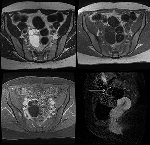






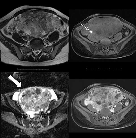


Adnexal masses often are indeterminate at ultrasound, the diagnostic imaging modality of choice in the workup of women with symptoms of such entities, and confirming ovarian etiology can be difficult. Additional diagnostic imaging is frequently indicated, as a common benign diagnosis may obviate the need for surgery, while prompt identification of malignant features requires intervention by subspecialist oncological surgeons.
Magnetic resonance imaging (MRI) is useful in the workup of complex adnexal masses. The modality has several problem-solving utilities that can be correlated with ultrasound findings, clinical history, physical examination and laboratory investigations when a diagnosis remains indeterminate. MR imaging also is more reproducible than sonography, and it can help determine additional diagnostic information, such as the presence of fat, blood products, fibrosis and enhancement pattern or diffusion restriction. DWI, for example, has been beneficial in differentiating benign from malignant tumors with high sensitivity.
Ovarian neoplasms range from benign to malignant and may be primary or secondary. Usually, they are classified by tissue of origin (surface epithelial, germ cell and sex-cord stromal) and metastatic (one secondary) (Table 1).
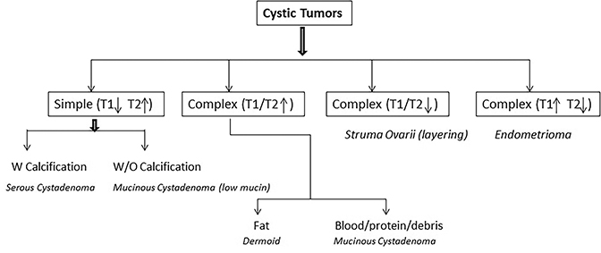
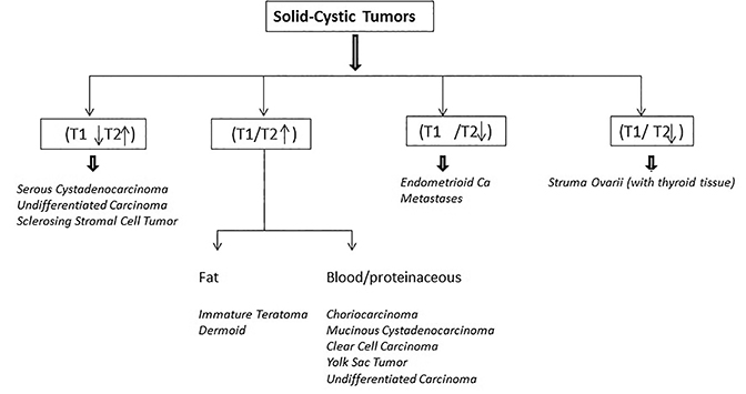
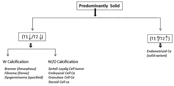
In this article, however, we classify ovarian masses into three main MR imaging categories: Cystic neoplasms (with septations), Complex neoplasms (solid-cystic) and Solid neoplasms (predominantly solid). These three categories are further subdivided according to specific MR imaging features, such as the presence of calcifications, fat, blood, proteinaceous content and signal intensities.
To the authors’ knowledge this is a novel approach. This MRI classification can help to narrow the differential diagnosis and add to the diagnostic confidence needed to guide the type and extent of definitive surgical management prior to a pathological tissue diagnosis. Indeed, the ultimate aim of this review is to help the radiologist to arrive at a narrow differential diagnosis and, in some cases, a specific diagnosis (Flowchart 1).
Cystic neoplasms
This category encompasses tumors predominantly cystic on morphology. These may or may not have internal septations, but they demonstrate no measurable solid component. MRI differentiating features include the presence of septations, fat, hemorrhage, proteinaceous content and calcification by typical T1 and T2 signal intensities. Fat appears as high signal on T1WI and T2WI with signal suppression on a fat-saturation sequence. Mucin is minimally to moderately bright on the T1WI (depending on concentration) but not brighter than fat on T1WI without fat suppression. Hemorrhage is also bright on the T1WI and T2WI with varied appearance (depending on the age). “T2 shading” is suggestive of chronic blood products (typical of endometriomas, Flowchart 2).
Simple cystic: T1 dark; T2 bright
These lesions are further divided into those with or without calcifications. Serous cystadenomas are benign epithelial tumors seen in the 4th to 5th decade. Generally smaller than mucinous tumors, these are unilocular or multilocular masses of simple fluid without papillary projections but containing psammomatous calcifications. On MRI they appear hypointense on T1WI, hyperintense on T2WI with mild enhancement of thin septations and no solid components (Figure 1).1
Complex cystic: T1 bright; T2 bright
These masses contain varying degrees of internal complexities such as fat, blood and proteinaceous content.
Mucinous cystadenomas are benign, mucin-containing epithelial tumors usually seen in women ages 30-50 years. They are larger than serous neoplasms, with a “honey-comb” of locules with varied signal intensities separated by thin septations and no solid components. Linear calcification is rarely noted. MRI shows a multilocular septated mass having simple or complex T1 and T2 signal, depending on mucin content.1 Loculi with watery mucin have lower signal intensity than loculi with thicker mucin. The locules appear bright on T2WI (Figure 2A).
Dermoid tumors are mature ovarian teratomas, slow-growing germ cell tumors that are almost always benign. They are small complex masses typically presenting around 20-30 years of age. The Rokitansky nodule is a projection into the cystic cavity and can contain bone, teeth or calcification, occasionally filled with fatty tissue. On MRI the sebaceous component has a very bright signal on T1WI and T2WI with suppression on the fat-saturation sequences (Figure 2B).1
Complex cystic: T1 dark; T2 dark
Struma ovarii are ovarian tumors composed of thyroid tissue, with 5-10% being malignant.2 Mostly occurring unilaterally in the reproductive years, they are multilocular, thick-walled masses with large, cystic spaces representing dilated thyroid follicles (filled with thyroglobulin and thyroid hormones). MRI shows a multiseptated, complex cystic mass. Some components of the cysts contain isointense to hypointense layering on T1WI and T2WI in dependent portions of the cyst, thought to represent thyroglobulin,2 a discerning feature for this lesion (Figure 3A). Avid enhancement of the thickened wall and septations correspond microscopically to thyroid tissue.2
Complex cystic: T1 bright; T2 dark
Endometriomas present in the reproductive age group and represent ectopic endometrial glands and stroma outside the uterus and a common cause of pelvic pain and infertility. MRI shows bright signal intensity on T1WI and dark signal intensity on T2WI, which helps to establish a diagnosis (Figure 3B). Endometriomas tend to have higher T1 and lower T2 signal intensities than hemorrhagic cysts. The greater degree of T1 and T2 shortening is attributable to higher protein concentration and viscosity. The lower T2 signal intensity of endometriomas has been described as “T2 shading.” No solid components or internal enhancement is seen.3
Solid-cystic neoplasms
These are complex cystic masses containing a solid enhancing component. Further differentiation is on the signal characteristics of T1- and T2-weighted images. Diffusion-weighted imaging and enhancement patterns offer additional clues. The solid malignant component (including thick septations) show restriction on DWI (compared to a benign entity). On dynamic contrast enhanced MRI, early enhancement patterns can help distinguish between benign, borderline and malignant/aggressive tumors (Flowchart 3).
Complex solid-cystic: T1 dark; T2 bright
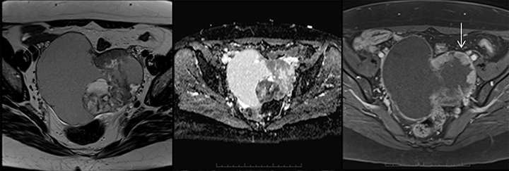
Serous cystadenocarcinomas, the malignant variant of the serous epithelial neoplasm, are among the high-grade cancers seen in older age groups (post-menopausal). Elevated CA125 and bilaterality are common. Smaller than mucinous neoplasms, these masses have solid components, papillary projections and psammomatous calcification. MRI usually shows these as T1 dark and T2 bright masses with DWI restriction and a strongly enhancing solid component.1 Rarely, tumoral hemorrhage may cause T1 bright signal (Figure 4A).
Undifferentiated carcinomas are seen in middle-aged women; these masses may be bilateral and show elevated tumor markers with an early propensity for metastatic disease and multiple histological cell types, suggesting an aggressive nature. MRI shows a variably sized, complex, T2 hyperintense, solid-cystic mass with occasional internal hemorrhage and necrosis. There is restriction on DWI, strong enhancement and metastatic peritoneal spread. Their aggressive, “wild” and early spread are helpful characteristics to diagnosing undifferentiated tumors.1 Given their varied imaging appearance these malignancies have overlapping features with serous cystadenocarcinoma, as both are epithelial tumors (Figure 4B)
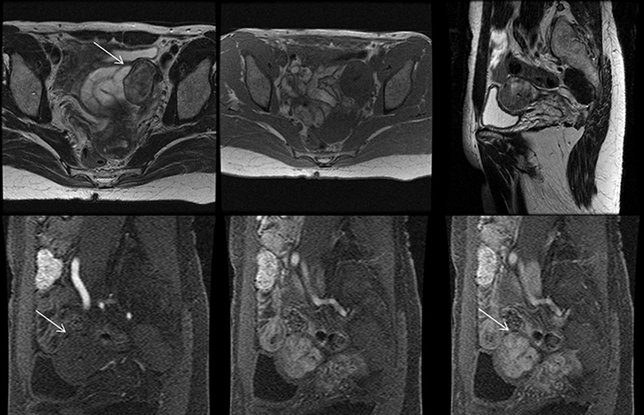
Sclerosing stromal tumors are rare tumors presenting in young women with menstrual cycle irregularity.4 Typically, these are seen as large masses with the solid component toward the periphery. MRI shows a T1 hypointense, T2 hyperintense mass with characteristic early peripheral enhancement on dynamic contrast-enhanced images and centripetal progression.1 The early enhancement reflects cellular areas with a prominent vascular network. The delayed enhancement in the inner portions represents collagenous hypocellular areas. This classic appearance is a diagnostic pearl specific to this tumor (Figure 5A).
Complex solid-cystic: T1 and T2 bright (fat, blood, proteinaceous)
Choriocarcinomas are among the rarest of the gonadal germ cell tumors associated with elevated β-HCG. Occurring in the reproductive age group, this mass may be misconstrued for an ectopic pregnancy. An enhancing, highly vascular complex solid-cystic mass with large signal voids and cavities is typically seen on MRI. These tumors are isointense to slightly hypointense on T1WI with scattered areas of hemorrhage, which when present are bright on T1- and T2-weighted sequences. Soft-tissue septae between the cavities show strong enhancement (Figure 5B).5
Immature teratomas peak between 15 and 20 years of age with elevated AFP and LDH and consist of mature and immature embryonic tissue. These tumors are large, encapsulated solid masses with cystic components, areas of necrosis and hemorrhage and fat, hair and sebaceous material. Typically MRI shows a large, irregular, solid-cystic mass containing coarse flecks of calcification and small, fat foci, necrosis and hemorrhage (Figure 6). These tumors grow rapidly and demonstrate capsular perforation.5
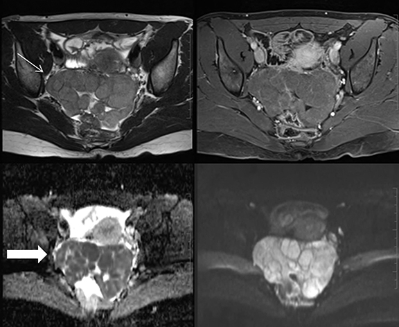
Mucinous cystadenocarcinomas are malignant mucinous neoplasms typically seen in an older population. Morphologically these are large, unilateral masses containing enhancing solid components and hemorrhage, occasionally with pseudomyxoma peritonei. Elevated CA 19-9 tumor marker is seen.6 MRI shows a large mass with variable complex (bright) signal on T1WI and T2WI. The presence of enhancing solid components and diffusion restriction confirms the diagnosis (Figure 7A). Serous and mucinous tumors show overlapping MRI features for malignancy; however, mucinous tumors are generally larger with a brighter signal on the T1WI.
Clear cell carcinomas are epithelial ovarian tumors, of which the benign and borderline variants are rare and usually malignant. Clear cell ovarian tumors peak at 55 years and can develop in a focus of endometriosis. They are usually solid-cystic with one or more polypoidal masses protruding into the lumen. MRI reveals a unilocular variable signal intensity cyst on T1WI (low to very high), bright signal on the T2WI and a few rounded, solid protrusions that restrict on DWI and show strong enhancement on the postcontrast sequence (Figure 7B).1
Yolk sac tumors, also known as endodermal sinus tumors, are large, encapsulated masses seen in young females (10-30 years) with elevated AFP. These neoplasms occasionally contain hemorrhage and necrosis. MRI shows a complex mass with variable (usually bright) signal on T1WI (depending on blood content), bright on T2WI and restriction on DWI (Figure 8). The bright dot sign is seen at contrast-enhanced CT and MRI as enhancing foci in the wall or solid components.5 These foci of enhancement are attributable to dilated vessels, considering the highly vascular nature of these tumors.
Complex solid-cystic: T1 bright; T2 dark (solid component, chronic blood products)
Endometrioid adenocarcinomas may be benign, borderline or malignant. Most common between 30 and 50 years of age, these tumors overlap considerably in morphology. Benign and borderline tumors are generally cystic and unilateral, with an excellent prognosis. Correlation with endometriosis has been documented for all three. The malignant variants may be cystic or predominantly solid, with a quarter being bilateral and associated with an endometrial carcinoma (regarded as an independent primary tumor). 7 MRI shows a complex solid-cystic enhancing mass typically hyperintense on the T1WI and a slightly lower signal on the T2WI (T2 shading) with an enhancing solid component (Figure 9).
Metastatic lesions in the ovaries most commonly originate from the colon and stomach. Krukenberg ovarian metastases contain mucin-secreting cells from the gastrointestinal tract. Imaging findings are nonspecific and often seen as a solid-cystic deposits (solid component is T2 dark) with strong enhancement postcontrast.1 High T1 signal on MRI may indicate mucin deposits. A history of a primary malignancy is beneficial information (Figure 10).
Complex solid-cystic: T1 and T2 dark
Struma ovarii, as described earlier, presents on MRI as a multilobulated complex mass with thick septae, multiple cysts and enhancing solid components (thyroid tissue). The cysts tend to appear hyperintense on T1WI and T2WI with hypointense layering in the dependent portion (Figure 3), the most discernable feature for diagnosis.2 The solid component shows strong enhancement.
Solid neoplasms
These tumors are predominantly solid on morphology with occasional scattered cystic foci. MRI classification pertains to appearances on T1WI, T2WI and enhancement (Flowchart 4).
Solid: T1 and T2 dark with calcification
Brenner tumors are incidental, usually benign epithelial tumors occurring in the 5th–7th decades, usually demonstrating calcifications. These can be solid-cystic (infrequently malignant) or mostly solid (common benign variant, incidentally discovered). MRI shows a T1 and T2 low signal mass with mild, delayed, postcontrast enhancement and amorphous calcification (Figure 11).3
Fibrous tumors, including fibromas, the commonest type, occur at all ages but most often in middle-aged women. MRI shows a solid, hypointense T1 dark and very T2 dark mass with none-to-faint enhancement (Figure 12). Dense calcification and scattered high signal areas indicate edema or cystic degeneration.4 High T1 suggests hemorrhagic infarction. An interface vessel between the uterus and adnexal masses is helpful to differentiate these masses from uterine leiomyomata.
Dysgerminomas are the ovarian counterparts of testicular seminomas; they are occasionally bilateral, most common in adolescence and associated with elevated β-HCG and LDH.8 Variably sized, these solid tumors contain speckled calcifications and foci of hemorrhage and necrosis. MRI features show a predominantly lobulated, solid, hypointense mass with mild enhancement postcontrast and restricted diffusion on DWI (Figure 13).
Solid: T1 and T2 dark without calcification
Sertoli-stromal tumors, also known as Leydig cell tumors, are rare, androgen-secreting, virilizing tumors in young women. They are usually unilateral and can be solid, solid-cystic, cystic or even papillary. MRI shows a moderately enhancing, solid,T2 hypointense mass with scattered intratumoral cysts that are bright on T2WI.9 The T2 low signal depends on the extent of fibrous stroma (Figure 14).
Embryonal cancer generally presents on MRI as a large, unilateral, predominantly solid mass containing cystic (mucoid-filled) spaces. Yolk sac tumors, immature teratomas and dysgerminomas are common components associated with embryonal carcinomas with all reported cases showing a mixed component. There is associated elevation of AFP and β-HCG. On MRI, these masses are isointense on T1WI, slightly hypointense on T2WI and show strong enhancement postcontrast.5 The cystic spaces are often hyperintense on T1WI and T2WI (hemorrhage) (Figure 15).
Granulosa cell cancer constitutes less than 5% of ovarian neoplasms (commonest malignant sex cord-stromal tumor) seen in postmenopausal women. A quarter can be associated with endometrial polyps, carcinoma or hyperplasia.1 There is a spectrum of imaging appearances, with heterogeneity caused by bleeding, infarction and fibrous degeneration. MRI shows a T1 isointense, T2 isointense-to- hypointense mass with cystic spaces (Figure 16A). There is mild-to-moderate postcontrast enhancement.4 The uncommon solid-cystic variant is bright on T2WI with a “spongy appearance.”
Steroid cell tumors, usually seen in the 5th or 6th decade, are a virilizing tumor associated, rarely, with Cushing’s syndrome. A third of these behave as clinically malignant. Usually small and unilateral, steroid cell tumors on MRI are isointense-to-hypointense on T1 and T2 with T1/T2 bright intratumoral cysts (lipid) and postcontrast enhancement (Figure 16B).4
Solid: T1 and T2 bright
Endometrioid tumors are predominantly solid, with a quarter being bilateral and associated with an endometrial carcinoma. MRI shows a solid T1 isointense and T2 hyperintense mass with postcontrast enhancement (Figure 17).7
Conclusion
MRI is an adjunctive imaging modality useful for characterizing indeterminate or complex ovarian masses after sonographic assessment. Although there are overlapping imaging features, MRI aids in making a specific diagnosis or narrowing differential diagnosis, thereby enabling more accurate clinical management. When malignant features are present, MRI can aid in planning definitive surgery.
References
- Jung SE, Lee JM, Rha SE, et al. CT and MR imaging of ovarian tumors with emphasis on differential diagnosis. RadioGraphics. 2002; 22(6):1305-1325.
- Matsuki M, Kaji Y, Matsuo M, et al . Struma ovarii: MR findings. BJR. 2000;73(865): 87-90.
- Valentini AL, Gui B, Miccò M, et al. Benign and suspicious ovarian masses – MR imaging criteria for characterization: pictorial review. J Oncol. 2012: 481806.
- Jung SE, Rha SE, Lee JM, et al. CT and MRI findings of sex cord–stromal tumor of the ovary. AJR Am J Roentgenol. 2005; 185(1): 207-215.
- Shaaban AM, Rezvani M, Elsayes KM, et al. Ovarian malignant germ cell tumors: cellular classification and clinical and imaging features. RadioGraphics. 2014: 34(3): 777-801.
- Kelly PJ, Archbold P, Price JH, et al. Serum CA19.9 levels are commonly elevated in primary ovarian mucinous tumours but cannot be used to predict the histological subtype. J Clin Pathol. 2010;63(2):169-175.
- Li HM, Qiang JW, Xia GL, et al. MRI for differentiating ovarian endometrioid adenocarcinoma from high-grade serous adenocarcinoma. J Ovarian Res. 2015; 8(26).
- Kitajima K, Hayashi M, Kuwata Y, et al. MRI appearances of ovarian Dysgerminoma. European Journal of Radiology Extra. 2007; 61(1):23–25.
- Cai SQ, Zhao SH, Qiang JW, et al. Ovarian Sertoli -Leydig cell tumors: MR findings and pathological correlation. J Ovarian Res. 2013;26(6):73.
Citation
J H, G L, U M. MRI classification and characterization of complex ovarian masses. Appl Radiol. 2017;(3):6-20.
March 4, 2017