Intraoperative 3D Imaging Improves Accuracy of Pedicle Screw Placement in Spine Surgery
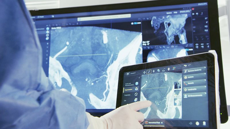
 Research presented at the American Academy of Orthopaedic Surgeons (AAOS) Annual Meeting by the Hospital for Special Surgery (HSS) found that intraoperative three-dimensional (3D) imaging was superior to two-dimensional radiographs in confirming the accuracy of pedicle screw placement during spine surgery.
Many spinal surgeries require the use of implants called pedicle screws to stabilize the spine. Precise positioning of these screws is critical for a successful outcome. “Two-dimensional biplanar radiographs [BPR] have been the gold standard to confirm pedicle screw placement in spine fusion surgery for many years. However, when utilizing BPR, there is the potential that a two-dimensional image alone will not properly demonstrate the successful placement of a screw,” said Darren Lebl, MD, a spine surgeon at HSS and principal investigator of the study. “As an alternative, three-dimensional intraoperative imaging systems are now available, offering improved visualization.”
Dr Lebl and colleagues set out to compare the accuracy of BPR versus 3D imaging when assessing intraoperative pedicle screw placement. “Our study is the first to compare the differences in intraoperative biplanar radiography and 3D imaging for pedicle screw accuracy in thoracic and lumbar cases using robotic technology,” Dr Lebl noted.
Investigators analyzed data from 103 patients who underwent spinal fusion by a single surgeon from 2019 to 2022. Pedicle screw placement was assessed with both intraoperative BPR and 3D imaging in each case.
“CT scans taken after surgery were compared to the findings of intraoperative BPR and 3D imaging to detect either false-positive or false-negative readings,” explained Fedan Avrumova, BS, an HSS clinical research coordinator who presented the study at the AAOS meeting. “False positive findings are instances when BPR imaging suggests the screw was not in an acceptable position, while in fact a more advanced 3D image (intraoperative 3D scan or postoperative CT scan) showed the screw to be in an acceptable position. Conversely, a false negative instance was when a BPR image led one to believe or looked as though the screw was in an acceptable position, when in fact a more advanced 3D image or post-operative CT scan showed that it was in fact not acceptable.”
Postoperative CT imaging revealed a clinically significant number of patients who had false-negative and false-positive screw placement readings on BPR. However, screw position shown on intraoperative 3D imaging was found to be much more accurate, Avrumova added.
“Based on our study, BPR imaging may lead one to think a screw is acceptable when in fact it is not, and also may miss many screws that are not in fact acceptable. In our study, it was approximately one percent of cases where this occurred. However, for surgeons and centers that implant hundreds and thousands of screws per year, this is going to result in a significant clinical impact for many people,” Dr Lebl noted. “Even one misplaced screw can have a significant impact for a patient, a surgeon, and a hospital system. Therefore, based on these findings, we suggest that for intraoperative confirmation of screw position 3D imaging may soon represent a new standard of care.”