Intraductal Papilloma in a Man
Case Summary
An adult man presented with a palpable, painful lump in the retroareolar region of the left breast, at the 3 o’clock position, present for 4 months. The patient denied a history of trauma, fever, nipple discharge, superficial erythema, and bruising. Further questioning revealed no similar symptoms in the past and no family history of breast cancer.
Imaging Findings
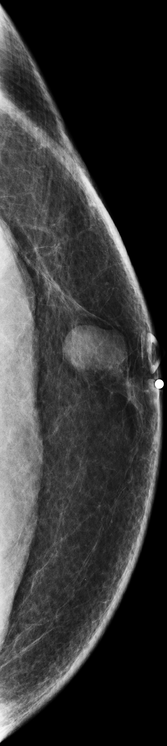
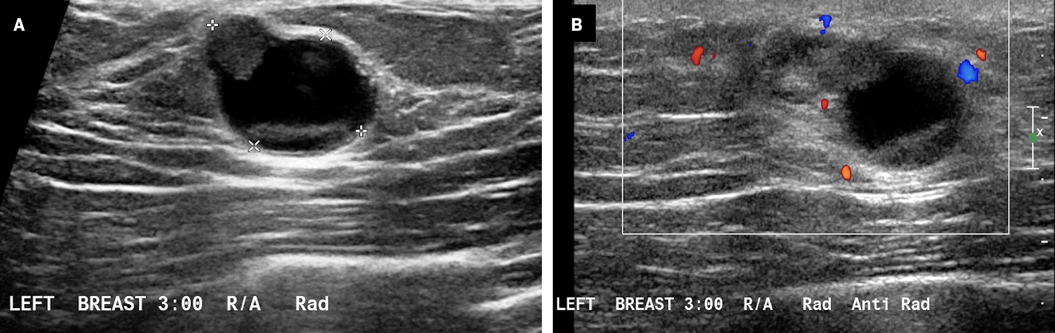
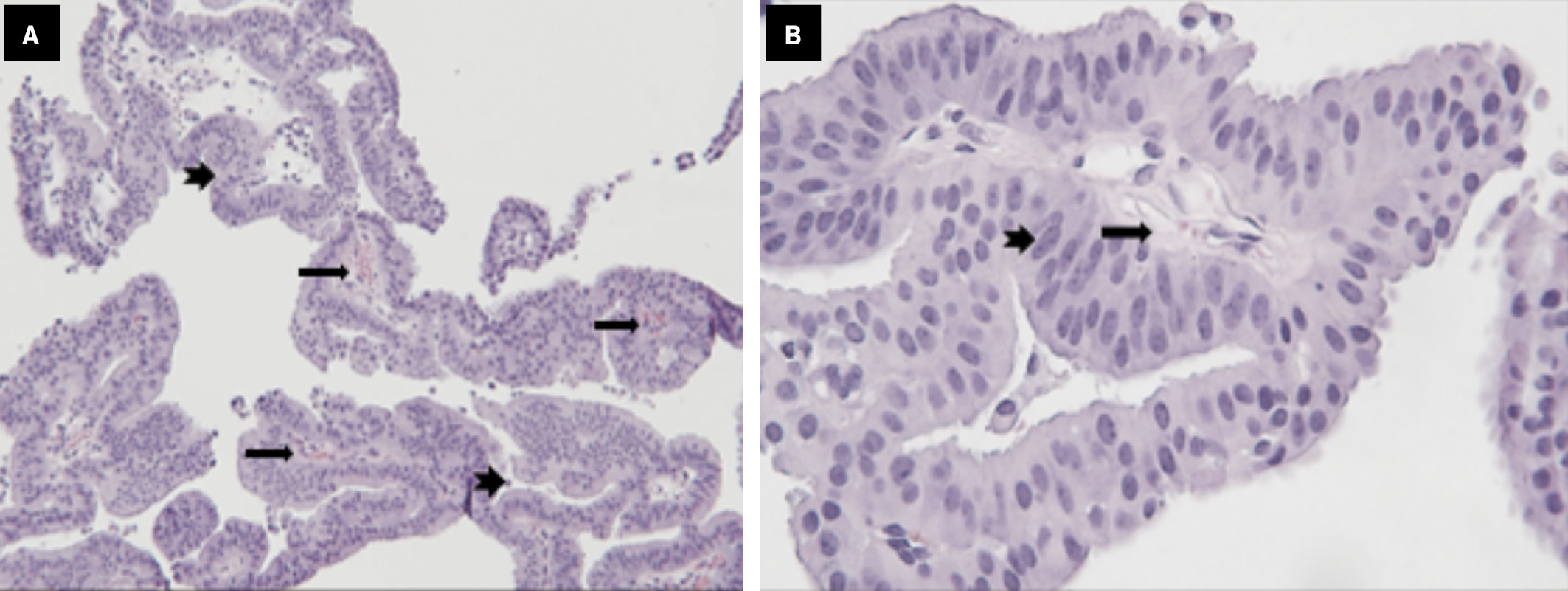
Diagnostic mammogram ( Figure 1 ) demonstrated a circumscribed, oval mass within the retroareolar left breast. Targeted breast US ( Figure 2 ) revealed an irregular, circumscribed, complex cystic and solid mass with posterior acoustic enhancement and peripheral Doppler flow. US-guided core needle biopsy was performed. Histopathologic analysis ( Figure 3 ) demonstrates highly fragmented papillae with fibrovascular cores lined by columnar cells with apocrine metaplasia.
Craniocaudal view of the left breast demonstrates an oval, circumscribed retroareolar mass without associated calcification or overlying skin thickening.
( A ) Targeted US of the left breast lesion, 3 o’clock position demonstrated a 1.5 × 1.1 cm irregular, circumscribed complex, predominantly anechoic, cystic lesion with solid components and posterior acoustic enhancement. ( B ) Peripheral Doppler flow and posterior acoustic enhancement are demonstrated.
( A ) Highly fragmented papillae with fibrovascular cores (arrows) lined by columnar cells with apocrine metaplasia (arrowheads) (H&E ×100). ( B ) The apocrine epithelium (arrowhead) lining the fibrovascular cores (arrow) shows uniform nuclei and prominent eosinophilic cytoplasm (apocrine metaplasia) (H&E ×400).
Diagnosis
Intraductal papilloma.
The differential diagnosis for breast lesions in men with similar presentation includes gynecomastia, invasive ductal carcinoma, ductal carcinoma in situ, abscess, lipoma, and epidermal inclusion cyst.
Discussion
The most common cause of bloody discharge from the female breast is intraductal papilloma, which is associated with 40-70% of cases of pathological nipple discharge in women.1 While intraductal papillomas are not uncommon in women, they are exceedingly rare in men. A literature review identified 15 published cases of histopathologically proven intraductal papilloma in male patients, aged 11-78 years between 1984 and 2020.1 - 5 Eleven patients presented with unilateral nipple discharge, which was bloody in a majority (73%) of those cases. Palpable masses were reported in 8 of the cases, and 10 cases without reported pain. A study by Zhong et al6 identified 117 cases of male-breast papillary lesions at a single medical center between 2000 and 2019. Five of these cases were pathology-proven, benign, intraductal papillomas.
Intraductal papillomas, although benign, are high-risk lesions with the potential for malignant transformation. One case in the literature demonstrated malignant transformation to invasive ductal carcinoma 3 years after diagnosis in a man who did not undergo surgical excision.4 Another case demonstrated an intraductal papilloma in a man with associated atypical ductal hyperplasia,7 which is also a high-risk feature. These were the only 2 cases in our review that reported associated pain.
The patient in our case is unique in that he presented with a painful, palpable lump, without nipple discharge. This combination of signs and symptoms was not reported in any of the cases we reviewed.
The histopathological features are diagnostic for this entity. Intraductal papillomas arise within ducts in central (solitary) or peripheral (multiple) locations.8 They are composed of papillae with fibrovascular cores, usually covered by both epithelial and myoepithelial layers.8 When the epithelium is apocrine, as in this case, the myoepithelial cells can be markedly diminished or completely lost.9, 10 These benign lesions can be associated with usual ductal hyperplasia, cuboidal to columnar cell changes, and apocrine change.8 Focal necrosis or hemorrhage may be present in larger lesions.
Conclusion
Given that intraductal papillomas are high-risk lesions, it is important to recognize that, although rare, they can occur in men. Our literature review identified 15-20 published cases of intraductal papilloma in men, the majority of who presented with painless, palpable lumps and nipple discharge. Our case highlights a unique presentation of intraductal papilloma in a man who presented with a painful lump, without associated nipple discharge.
References
Citation
Schwartz SD, Choudhury MS, Jacques SM, Singh E.Intraductal Papilloma in a Man. Appl Radiol. 2025; (1):29-30.
doi:10.37549/AR-D-24-0053
February 10, 2025