Graft versus host disease
Images


CASE SUMMARY
A 6-year-old boy presented with rash, vomiting, and diarrhea. His medical history was significant for pre-B cell acute lymphoblastic leukemia status post-allogenic hematopoietic stem cell transplant 55 days previous.
IMAGING FINDINGS
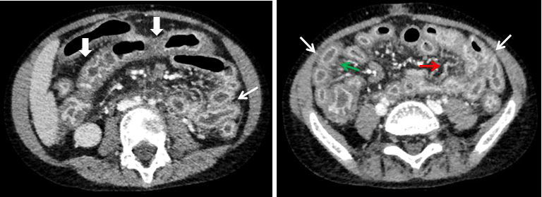
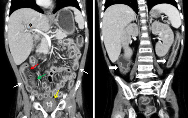
Axial and coronal CT images of the abdomen and pelvis with intravenous contrast (Figures 1-3) demonstrated diffuse mural thickening and excessive mucosal enhancement of the small and large bowel, with associated engorgement of the vasa recta and mesenteric stranding. The bowel was diffusely fluid filled, consistent with patient’s history of diarrhea.
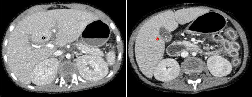
The liver was diffusely enlarged, consistent with hepatomegaly. There was diffuse intrahepatic periportal edema, which was causing mass effect and narrowing of the portal veins. Gallbladder abnormalities included mural thickening, excessive enhancement, cholelithiasis, intraluminal sludge and pericholestic fluid.
There was diffuse mural thickening and abnormal mucosal enhancement of the bladder. Patient had known BK virus-associated hemorrhagic cystitis.
DIAGNOSIS
Graft versus Host Disease (with intestinal and hepatic involvement).
Differential diagnosis includes neutropenic enterocolitis, clostridium difficile colitis, and viral enterocolitis.
DISCUSSION
Graft versus host disease (GVHD) continues to be a major cause of morbidity and mortality in patients who have received allogenic hematopoietic stem cell transplantation (HSCT). GVHD is a multi-organ disease secondary to activated donor immune cells attacking host tissue, and it occurs in both acute and chronic forms. 1,2 The main target organs involved in GVHD are the skin, gastrointestinal tract, and liver.1 Clinical manifestations may include rash, crampy abdominal pain, nausea/vomiting, voluminous diarrhea, and elevated bilirubin and/or liver enzymes. 1,2 The most significant risk factor for developing GVHD is HLA mismatch, with a greater degree of mismatch corresponding to a higher likelihood of developing GVHD. 2 Typically, symptom onset is before 100 days after the stem cell transplant during the donor engraftment stage.2 The modified Glucksberg criteria, which account for the number and extent of target organ involvement, are commonly used to assess the severity of acute GVHD.1,2 Severity ranges from mild, skin-only involvement (grade I) to multi-organ disease with severe skin and/or severe hepatic involvement (grade IV).1,2 Depending on the source and degree of HLA mismatch, the incidence of grade II-IV acute GVHD in the pediatric population ranges between 19%-85%.2
Typical CT findings of GI-acute GVHD include moderate bowel-wall thickening, mucosal hyperenhancement, bowel dilatation, fluid-filled small bowel, vasa recta engorgement, and mesenteric stranding.4 Any portion of the GI tract can be involved, from the esophagus to the rectum, and involvement may be discontinuous. Distribution may be helpful in distinguishing GVHD from other common causes of bowel inflammation in the HSCT patient. Neutropenic enterocolitis is often limited to the ileocecal region, with Clostridium difficile colitis manifesting as a pancolitis.
CT findings with acute hepatic GVHD are variable and nonspecific. Hepatomegaly is almost universally present, with other possible findings including splenomegaly, ascites, or periportal edema.3 Interestingly, biliary tract abnormalities, including abnormal biliary tract enhancement, dilatation of the common bile duct, pericholecystic fluid, gallbladder wall thickening, and biliary sludge, are commonly seen coexisting with acute GI GVHD and are not necessarily indicative of acute hepatic GVHD. 4
Although mild acute GVHD (grade I) may be treated conservatively, systemic glucocorticoid steroids are the first line treatment for moderate and severe acute GVHD (grades II-IV).1 Approximately 50% of patients respond to the initial steroid therapy. Patients with higher grades of GVHD, and those who received HLA-mismatched donor transplants, are less likely to respond.2 Patients with acute GVHD are at increased risk of developing chronic GVHD. Long-term survivability rates are inversely related to the grade of acute GVHD, with approximately 30% of patients with grade III GVHD achieving long-term survival, versus 5% of patients with grade IV GVHD.2
CONCLUSION
Graft versus host disease is a potentially devastating complication following allogenic hematopoietic stem cell transplantation. Unfortunately, it is difficult to distinguish the clinical manifestations of GVHD from other potential complications following HSCT, which typically require a drastically different treatment plan. Radiologists can serve an important role in the correctly diagnosing GVHD early in the disease course by recognizing its classic CT imaging features of diffuse bowel-wall thickening with mucosal hyperenhancement, bowel dilatation, fluid-filled small bowel, vasa recta engorgement, and mesenteric stranding.
REFERENCES
- Carpenter P, MacMillan M. Management of acute graft versus host disease in children. Pediat Clin North Am. 2010. 57:273-295.
- Jacobsohn D Acute graft-versus-host disease. Bone Marrow Transplantation. 2008; 41:215-221.
- Erturk S, Mortele K, Binkert C et al. CT features of hepatic venooclusive disease and hepatic graft-versus-host disease in patients after hematopoietic stem cell transplantation. AJR.2006; 186: 1497-1501.
- Mahgerefteh S, Sosna J, Bogot N et al. Radiologic imaging and intervention for gastrointestinal and hepatic complications of hematopoietic stem cell transplatation. Radiology. 2011; 258:660-671
Citation
JC W, SA J, AJ T, R T. Graft versus host disease. Appl Radiol. 2017;(12):.
December 13, 2017