Gamekeeper’s thumb (Skier’s thumb)
Images

Gamekeeper’s thumb (Skier’s thumb)
Findings
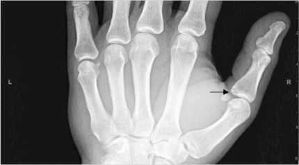
AP and lateral radiographs showed a fracture at the base of the proximal phalanx of the thumb (Figure 1). A small osseous fragment was noted on the ulnar side of the first metacarpophalangeal (MCP) joint (Figure 2). A concave defect was noted in the base of the proximal phalanx of the thumb, consistent with a “donor site.” This location is the attachment site of the ulnar collateral ligament, one of the 2 collateral ligaments of the first MCP joint.
Discussion
Gamekeeper’s thumb is a common injury and is used to describe an acute or chronic injury of the ulnar collateral ligament of the first metacarpophalangeal joint. The term was initially used to describe chronic occupational injuries to the ulnar collateral ligament to gamekeepers in Scotland because of their methods used to kill rabbits.1 The injury occurred in gamekeepers as they sacrificed rabbits by breaking their necks with their thumbs and index fingers. Acute injuries now tend to be more common among skiers and may constitute up to 50% of hand injuries in these athletes. It may also be seen in patients with rheumatoid arthritis and suffering from a motor vehicle accident and other sports injuries resulting from a fall onto an outstretched hand with an abducted thumb.
The ulnar collateral ligament is a short ligament that originates from the metacarpal head and inserts onto the medial aspect and base of the proximal phalanx of the thumb. The MCP joint chiefly provides flexion and extension movements. Stability to the joint during activity is provided primarily by the intrinsic and extrinsic muscles of the thumb. The proper collateral ligament, the accessory collateral ligament, the palmar plate, and, to some extent, the dorsal capsule also help to stabilize the MCP joint. The adductors of the thumb insert onto the extensor expansion through its aponeurosis, which lies superficial to the ulnar collateral ligament.
Most injuries of the thumb are ligamentous injuries, usually involving an avulsion of the ulnar collateral ligament from the base of the proximal phalanx. The mechanism of the injury involves an acute valgus stress on the MCP joint during a fall on an outstretched hand.Occasionally, an avulsion of the bone at the base of the proximal phalanx may result in a gamekeeper’s fracture.
Clinical evaluation and radiographs can be used to make the diagnosis. Prior to any manipulation of the thumb, standard anteroposterior,lateral, and oblique radiographs must be obtained to exclude metacarpal fractures and gamekeeper’s fractures. The finding of 3 mm of volar subluxation of the phalanx on the metacarpal is suggestive of complete UCL rupture and instability. Radial deviation of >40° in extension and >20° in flexion indicates instability. Stress radiographs obtained with the thumb in the flexed and extended positions and with valgus stress at the MCP joint can help in determining the instability of partial tears of the UCL.2
A grade I or II sprain without evidence of joint laxity is treated conservatively. Nonsurgical treatment by immobilizing the thumb in aspica-type cast for 4 weeks can be considered in partial tears of the ulnar collateral ligament. A grade III sprain, as a result of a complete tear with laxity at the MCP joint and angulation >30° to 35°3 results in chronic laxity, pain, loss of grip strength, and eventual degenerative osteoarthropathy.4 Complete UCL tears require surgical intervention. Gamekeeper’s fractures are usually treated conservatively, but those involving >30% of the joint surface and those that are malrotated and/or displaced should not be manipulated. Those fractures are indications for surgical intervention.2
A Stener lesion5 could also result from this injury. The prevalence of this is variable and in one series was reported at 27%.4 It is a result of the torn end of the ulnar collateral ligament being displaced and coming to lie superficial to the adductor pollicis aponeurosis. This lesion also can be associated with gamekeeper’s fracture, which can be subtle or obvious. Stener lesions may produce a lump over the medial aspect of the MCP joint, but otherwise they can be difficult to diagnose clinically. A lump or mass over the ulnar aspect of the MCP joint should raise suspicion for a Stener lesion if no fracture is noted on plain radiographs. Plain films, stress views, and arthrograms, which have been used to diagnose ruptures of the ulnar collateral ligament, do not provide sufficiently accurate images in diagnosing Stener lesions.6,7,8,9
The UCL no longer contacts its area of insertion and due to the interposition of the aponeurosis between the proximal and distal ends of the torn ligament, healing in these lesions can be impaired. These lesions are initially treated conservatively for 6 weeks. If instability still persists despite conservative treatment, surgery is undertaken. However, if surgery is undertaken 3 weeks after the initial injury, intervention is more difficult and the results are suboptimal.10 Complete tears repaired after 3 weeks tend to have an increased incidence of weakness and pain on pinch grasp. For this reason, early surgical intervention is generally undertaken for all cases of ruptured ulnar collateral ligaments.
Studies have shown the utility of magnetic resonance (MR) imaging in establishing the integrity of the ulnar collateral ligament.4,11,12 MR findings can be used after rupture of the ulnar collateral ligament to distinguish between a Stener lesion and a nondisplaced or minimally retracted tear. This could further help to distinguish surgical patients from those who can be treated nonsurgically.4 Early and accurate diagnosis would help to avoid unnecessary surgeries and be cost effective, thus justifying the cost of MR imaging.4
Ultrasonography (US) is also considered safe and accurate and is currently being used for the direct evaluation of the UCL of the thumb. USis a more dynamic and less time-consuming test than MR and may be easier to perform. Other disorders, such as tenosynovitis, tendon tears, and articular pathologic conditions can also be diagnosed with US. In this context, the authors suggest that US is an underused tool as it is potentially an adjunct to the clinical examination in the appropriate setting.13
Conclusion
Gamekeeper’s thumb is a common skiing injury. Early and accurate diagnosis of this injury and its complications can result in better patient outcomes and decreased long-term morbidity.
- Campbell CS. Gamekeeper’s thumb. J Bone Joint Surgery. 1955;37-B:148-149.
- Hannibal M. Orthopedic surgery for gamekeeper’s thumb. Coauthor(s): Roger D. Medscape. http://emedicine.medscape.com/article/1239413-overview#a0103. 2004.
- Posner MA, Retailaud J. Metacarpophalangeal joint injuries of the thumb. Hand Clinics. 1992;8:713-732.
- Hinke DH, Erickson SJ, Chamoy L, Timins ME. Ulnar collateral ligament of the thumb: MR findings in cadavers, volunteers, and patients with ligamentous injury (gamekeeper’s thumb), AJR Am J Roentgenol. 163:1431-1434.
- Stener B. Displacement of the ruptured ulnar collateral ligament at the metacarpophalangeal joint of the thumb. J Bone Surg. 1977;59-A: 519-524.
- Downey EF Jr, Curtis DJ. Patient-induced stress test of the first metacarpophalangeal joint: A radiographic assessment of collateral ligament injuries. Radiology.1986;158:679-683.
- Resnick D, Danzig LA. Arthrographic evaluation of injuries of the first metacarpophalangeal joint: gamekeeper’s thumb. AJR Am J of Roentgenol. 1976:126:1046-1052.
- Stener B, Stener I. Shearing fractures associated with rupture of the ulnar collateral ligament of the metacarpophalangeal joint of thumb. Injury. 1969;1:12-16.
- Stener B. Skeletal injuries associated with rupture of the ulnar collateral ligament of the metacarpophalangeal joint of the thumb. Acta Chir Scand. 1963;125:583-586.
- Arnold DM, Cooney W, Wood M. Surgical management of chronic ulnar collateral ligament insufficiency of the thumb metacarpophalangeal joint. Orthopedic Review. 1992;21;583-588.
- Louis DS, Buckwaller KA. Magnetic resonanace imaging of the collateral ligaments of the thumb. J of Hand Surg (Am). 1989;14A:739-741.
- Spaeth JA, Abrams RA, Bock GW, et al. Gamekeeper’s thumb: Differentiation of non-displaced and displaced tears of the ulnar collateral ligament with MR imaging. Radiology. 1993;188:553-556.
- Ebrahim Farhad S, Maeseneer Michel De, Jager Tjeerd, et al. US diagnosis of UCL tears of the thumb and Stener lesions: Technique, pattern-based approach, and differential diagnosis. Radiographics. 2006; 26:1007-1020.