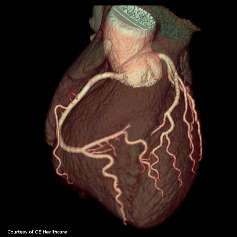
Deep-Learning Segmentation Reduces Postprocessing Time for CT of Chronic Total Occlusion
 A study published in Radiology reports that a deep-learning model to automate segmentation and reconstruction for CT of chronic total occlusions (CTO) considerably reduced postprocessing time for quantification. The tool was also found to correlate and be in agreeing in the anatomic assessment of occlusion features.
Coronary CT angiography (CCTA) guides coronary stenosis diagnosis and provides valuable information on CTO characteristics, including lesion length, calcium burden, stump morphology and tortuosity. However, conventional reconstruction of CCTA images for CTO is time consuming because commercially available software cannot automate nonopacified occluded segments, requiring manual reconstruction.
The researchers from Shanghai General Hospital aimed to develop a DL model to automate CTO reconstruction and compared to conventional, manual quantification. They used a CT training set from 6,600 patients, of whom 582 had CTO, and a validation set of 1,962 patients, of whom 208 had CTO. An external test set of 211 patients with 240 CTO lesions was used for validation. Consistency and validation between the DL model and conventional, manual reconstruction were compared using the intraclass correlation coefficient, Cohen Kappa coefficient, and Bland-Altman plot. The predictive values of CT-derived Multicenter CTO Registry of Japan (J-CTO) score for revascularization success were evaluated.
The authors report the DL model was successful in 95% of the lesions (228) without manual editing compared to 48% (116) of the lesions with the conventional reconstruction protocol. Time saved using he DL method was a mean of 335 seconds (mean, 121 seconds ± 20 [SD] vs 456 seconds ± 68; P< .001). In their conclusion, the authors wrote that in addition to significantly reducing postprocessing time for chronic total occlusion (CTO) quantification on coronary CT angiographic images, the anatomic assessment of occlusion features based on the DL model had excellent correlation and agreement when compared with manual reconstruction anatomic assessment.