Computed tomography of the acute abdomen
Images
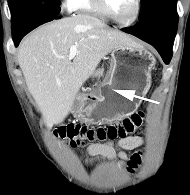
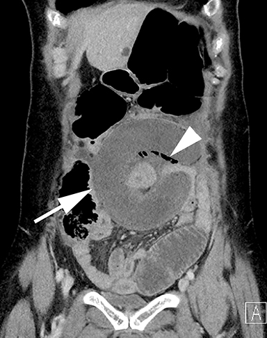
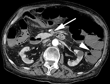
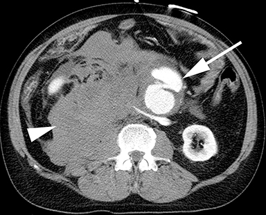
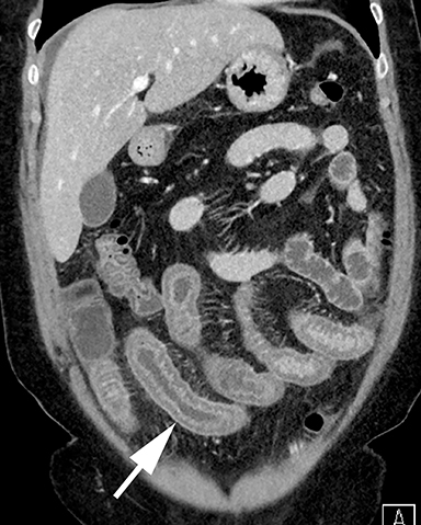
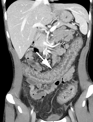
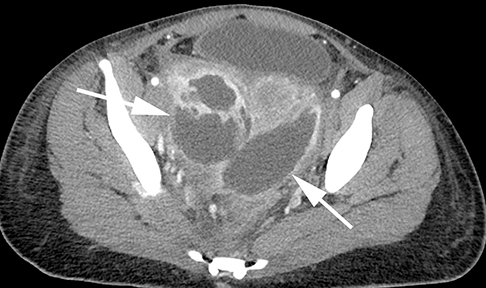
Acute abdominal pain is a common symptom for seeking urgent medical evaluation. The term ‘acute abdomen’ has historically referred to patients needing immediate surgical intervention, but it has broadened to include any patient experiencing acute pain to a degree that requires medical evaluation. The causes of acute abdomen are numerous and span the medical and surgical spectrum, with many etiologies identifiable using medical imaging. Indeed, radiology in the emergency department and the acute setting plays an important role in the diagnosis and workup of these patients, with imaging adding value to patient care.1
This review will focus on common causes of the acute abdomen that are detected on imaging, particularly computed tomography (CT). Typical imaging findings as well as pitfalls and complications will be discussed.
CT Technique
While protocols will vary by institution, scanning is generally performed from the diaphragm through the pubic symphysis. Images are reconstructed as contiguous 2.5-5 mm axial images and reformatted as 2-3 mm coronal and sagittal images. Depending on the scanner, there are options for dose reduction with automated tube voltage and current modulation. Intravenous (IV) contrast material is essential, typically with a single acquisition in the portal venous phase that can be obtained using a fixed scan delay or bolus tracking with automated triggering.
At our institution, positive enteric contrast (iodinated water soluble contrast) is rarely utilized in the emergency department (ED). By not utilizing enteric contrast, studies have shown improved patient throughput while retaining high accuracy.2,3 Positive enteric contrast can be useful for select indications, including assessing for fistulae, enteric leak and mesenteric abscess; however, early intestinal ischemia and GI bleeding can be masked by enteric contrast. We find that with thin-image reconstruction and multiplanar reformatted imaging, the lack of enteric contrast does not affect our accuracy or confidence.
Dual energy CT (DECT), in which two different photon energies are used in a single acquisition, allows for the creation of a virtual non-contrast dataset. While there is potential for dual energy CT to allow characterization of incidental adrenal and renal lesions using a single post-contrast acquisition, small renal calculi can be missed on virtual noncontrast datasets, and in the setting of acute abdominal pain, the utility of DECT is unclear and research is ongoing.4-6
Differential diagnosis
The differential diagnosis for the acute abdomen is broad. For patients with right upper quadrant pain, cholecystitis should be considered. Appendicitis and gynecologic etiologies commonly present with right lower quadrant or pelvic pain. Diverticulitis most commonly affects the sigmoid colon and patients often present with left lower quadrant pain. Epigastric pain may suggest acute pancreatitis or peptic ulcer disease, while flank pain should suggest urinary tract pathology. Generalized abdominal pain should raise the suspicion for bowel obstruction and mesenteric ischemia. While a quadrant-based differential diagnosis may be useful, the specific abnormalities may present with nonspecific symptoms, and a careful search pattern on CT imaging is required.
Acute appendicitis
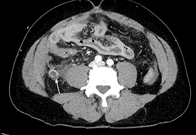
Acute appendicitis is a common acute surgical condition, with an incidence of 100 per 100,000 person-years in North America.7 Signs and symptoms can include right lower quadrant pain, nausea/vomiting, loss of appetite, fever, and leukocytosis. Imaging findings include a dilated appendiceal lumen, wall thickening, mural hyperenhancement, and periappendiceal inflammatory changes (Figure 1). Mimics of appendicitis can include mucocele (dilated, fluid filled appendix without periappendiceal inflammatory change), inflammatory bowel disease, acute diverticulitis (ileal or colonic), carcinoma, epiploic appendagitis, and gyncecologic abnormalities.8 Missed appendicitis may be caused by an unusual location of the appendix (right upper quadrant, left hemiabdomen), localized inflammation of the tip of the appendix (tip appendicitis), inflamed appendiceal stump following appendectomy, falsely-reassuring presence of intraluminal gas, or a separate concurrent process causing the reader to overlook the appendix.8,9
Complications of untreated appendicitis can include perforation, abscess formation, and septic seeding of the portomesenteric venous system. While antibiotic therapy alone is a validated option for the treatment of acute appendicitis, most providers and patients prefer definitive surgical management.10,11
Acute cholecystitis
Acute cholecystitis is a common cause of the acute abdomen and patients most often present with right upper quadrant pain.12-14 The disease can be either calculous or acalculous, with the former representing the large majority of cases.
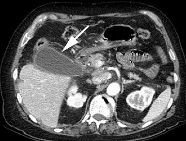
Acute cholecystitis is a clinical diagnosis that requires appropriate correlation between history and physical examination, laboratory studies, and radiologic examination. Cholecystitis can be demonstrated using ultrasound, CT, MRI, or nuclear scintigraphy, though the ACR appropriateness criteria lists ultrasound as the most appropriate initial imaging in a patient with suspected acute cholecystitis.15 CT findings of acute cholecystitis include wall thickening and luminal distention, though pericholecystic inflammatory change is often a central finding (Figure 2). CT also offers a complete picture of the surrounding tissues and allows for visualization of complications of acute cholecystitis such as perforation, emphysematous cholecystitis, hemorrhage, gallstone ileus, and postoperative complications.13 Gallstones can be seen on both CT and ultrasound, though ultrasound is more sensitive.16
In addition to common findings, a number of CT signs have been described that suggest the diagnosis. The tensile gallbladder fundus sign, defined as the absence of gallbladder fundal flattening by the abdominal wall due to increased gallbladder pressures, has been shown to be useful, particularly early in the presentation.17 Similarly, the pope’s hat sign has been recently described as a thin crescent of low density between the gallbladder and liver and may also improve diagnostic performance in early acute cholecystitis.18
Once the diagnosis is made, treatment options typically include cholecystectomy or percutaneous cholecystostomy tube placement. CT scans may be obtained post treatment to assess for postoperative complications or cholecystostomy tube position. Biliary scintigraphy is also used when bile leaks are suspected.
Acute diverticulitis
Approximately 10-20% of the millions of Americans with diverticulosis will develop acute diverticulitis, caused by impaction of a diverticulum and development of focal inflammation. The left colon is affected in 90% of cases and patients commonly present with left sided or left lower quadrant pain.19 Diverticula; however, can occur throughout the GI tract and cecal, ascending colonic, and small bowel diverticulitis are all possible.
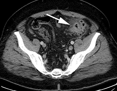
CT is excellent at portraying findings of both uncomplicated and complicated diverticulitis. For the former, findings with a high sensitivity include colonic wall thickening and pericolonic inflammatory change (Figure 3).20 Diagnostic confidence can be increased by finding a culprit diverticulum, i.e. a single outpouching that appears acutely inflamed or at the epicenter of the surrounding stranding. Adjacent fascial thickening can also increase specificity.21 Complications of diverticulitis include perforation, intramural abscess, and pericolonic abscess. Perforation may result in localized foci of pneumoperitoneum, or may less commonly cause diffuse or large volume free air. If untreated or longstanding, chronic diverticulitis may lead to complications including colovesical and colocolonic fistulas, intramural tracts, and chronic fluid collections.
Various pitfalls and mimics exist when imaging acute diverticulitis. The most important mimic of diverticulitis is colon cancer, which can also cause wall thickening, fistulae, and surrounding inflammatory change. Cancers tend to be associated with pericolonic lymph nodes, intraluminal masses, asymmetric wall thickening, and shorter segments of involvement, though considerable overlap in findings exists. While some international guidelines recommend colonoscopy after every case of diverticulitis, a growing body of literature has suggested that colonoscopy is not routinely necessary following uncomplicated acute diverticulitis.22,23
Treatment of uncomplicated diverticulitis is often conservative and comprised of antibiotics and dietary change. Complicated cases may require percutaneous drainage of fluid collections or surgery for free intraperitoneal gas, and recurrent or chronic cases may eventually be referred for resection of the affected colon.
Acute pancreatitis
Acute pancreatitis is a common etiology of acute abdominal pain in patients presenting to the ED. The clinical diagnosis of acute pancreatitis requires two of three features: 1) epigastric pain 2) elevated serum lipase to three times normal and 3) characteristic findings on imaging.24 In the early phase of acute pancreatitis (first week), treatment is supportive and severity is determined by the presence or absence of organ failure caused by the systemic inflammatory response syndrome. Treatment during the late phase (beyond the first week) is determined by the presence of symptoms or complications of pancreatitis and is often based on imaging findings including pancreatic necrosis, fluid collections, and pseudoaneurysms.24
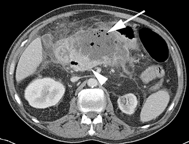
The Revised Atlanta Classification describes two morphologic appearances of acute pancreatitis: interstitial and necrotizing.24 Interstitial edematous pancreatitis (IEP) appears as focal or diffuse enlargement of the pancreas with homogeneous or slightly heterogeneous enhancement and peripancreatic soft tissue stranding and fluid. Necrotizing pancreatitis includes both pancreatic parenchymal and peripancreatic necrosis. Pancreatic parenchymal necrosis can be diagnosed when portions of the pancreas lack enhancement and should be described as <30%, 30-50%, or >50% of the gland. Peripancreatic necrosis can be distinguished by peripancreatic fluid that contains non-liquid/fat components (Figure 4).
Interstitial edematous and necrotizing pancreatitis can be complicated by both acute (< 4 weeks) and chronic (> 4 weeks) fluid collections that may be sterile or infected (Figure 4). Acute fluid collections in the setting of IEP rarely become infected and rarely require percutaneous or surgical intervention. Pseudocysts are chronic fluid collections in the setting of IEP which also rarely become infected but may require endoscopic or percutaneous drainage if symptomatic. Acute necrotic collections and walled off necrosis may require surgical, endoscopic or percutaneous drainage depending on a patient’s symptoms and do require therapy if infected.24
Pseudoaneurysms or active hemorrhage can be diagnosed at CT imaging and are commonly treated with transarterial embolization. Venous thrombosis is common in the setting of severe acute pancreatitis, but treatment is largely supportive.
Peptic ulcer disease
Peptic ulcer disease (PUD) is most commonly caused by Helicobacter pylori bacteria and non-steroidal anti-inflammatory medication. Common complications of PUD include GI bleeding and perforation, with gastric outlet obstruction and fistulization occurring less frequently. There are direct signs (focal outpouching and mucosal enhancement defects) and indirect signs (edema, wall thickening, and adjacent soft tissue stranding) of ulcer disease (Figure 5).25 Inadequate luminal distention often limits evaluation of the stomach on CT and is a potential pitfall for identifying wall thickening accurately.26 While most ulcers are occult at CT, evaluation for direct and indirect signs and use of multiplanar reformatted imaging can improve sensitivity.27 Duodenal ulcers and marginal ulcers at a gastrojejunostomy are more difficult to identify and a high index of suspicion is required. Perforation related to PUD can be readily detected using CT; gastric ulcers will typically cause pneumoperitoneum, while duodenal ulcers may cause intraperitoneal or retroperitoneal air depending on the site of perforation.
Small-bowel obstruction
Patients with small-bowel obstruction (SBO) generally present with nausea and vomiting with a distended abdomen. Postoperative adhesions are the most common cause of bowel obstruction, followed by hernias and malignancy. Abdominal radiography is historically the first imaging test for patients with suspected SBO; however, CT imaging can add value for clinicians by revealing SBO etiology and severity.28 At CT, proximal loops of small bowel will be dilated (> 2.5 cm) and there will be a transition to normal caliber or decompressed loops of small bowel at the site of obstruction.29 In addition, bowel may contain gas-mottled material proximal to the site of obstruction, known as the “small bowel feces” sign. Patients with uncomplicated bowel obstruction are often treated conservatively with enteric tube decompression and bowel rest.
Complicated small-bowel obstructions include strangulation with ischemia, closed loop obstructions, and internal hernias. These entities often require urgent surgical evaluation, and CT can help with prompt diagnoses. Findings of early bowel ischemia include wall thickening, abnormal enhancement (hypo- or hyperenhancement), and mesenteric edema, with pneumatosis intestinalis, portal and mesenteric venous gas, and pneumoperitoneum seen in later stages.29
Closed loop obstruction occurs when a segment of bowel is obstructed at two sites, isolating the obstructed segment and putting it at high risk of ischemia. A closed loop can be caused by a single adhesive band or a mesenteric twist/volvulus.30 The CT diagnosis of a closed loop obstruction is complex and the associated imaging findings of a C- or U-shaped obstructed segment, a “beak” sign, a mesenteric “swirl” sign, and interloop fluid/edema have variable sensitivity (Figure 6).31
Internal hernias can be congenital or acquired but are increasingly seen in the setting of Roux-en-Y gastric bypass. Several signs have been described including swirled mesenteric vessels, convergence of the mesenteric vessels as they pass through the hernia, and clustered loops of small bowel. The combination of the mesenteric swirl and convergence of the mesenteric vessels has been found to be the best indicator of an internal hernia.32 Internal hernias can be difficult to identify, but clinical suspicion and the use of multiplanar reformatted images can help.
Vascular: Mesenteric ischemia/AAA
Most acute abdominal pain is not related to a vascular etiology, yet vascular causes of abdominal pain can be associated with high morbidity and mortality. The most common vascular causes of acute abdominal pain include mesenteric ischemia and abdominal aortic aneurysm (AAA).33
Mesenteric ischemia can be occlusive (70-80%) or non-occlusive (20-30%). In the former, there is occlusion of the superior mesenteric artery or vein (Figure 7) leading to bowel ischemia from either insufficient inflow or venous hypertension and congestion. Non-occlusive mesenteric ischemia is seen in the setting of systemic hypotension and decreased mesenteric perfusion. At CT, the mesenteric vasculature should be assessed for patency, and one should search for signs of bowel ischemia, including dilatation (ileus), bowel wall thickening, and hypo- or hyper-enhancement. Bowel wall thinning is occasionally seen and may indicate impending perforation. Pneumatosis and mesenteric/portal venous gas are generally late findings and indicate necrosis.20
AAA is readily visualized on CT. The presence of high-density fluid in the retroperitoneum should raise concern for rupture (Figure 8). Findings suggesting impending rupture include increased aneurysm diameter (if prior imaging is available), acute intramural hematoma, perianeurysmal soft tissue stranding, and draping of the aorta over the spine.34 These findings warrant emergent surgical or endovascular intervention.
Non-ischemic bowel wall thickening
Small bowel and colonic wall thickening are nonspecific findings that may be the result of ischemic, infectious, or inflammatory etiologies. Normal wall thickness is less than 3 mm; however, thickness depends on distention. Non-distended small bowel will appear thicker and will enhance more than well-distended small bowel. In addition, the jejunum appears thicker and enhances more than the ileum due to increased mucosal surface area. With tube current modulation (80-100 kVp), the bowel wall can appear hyperemic following IV contrast, a result of obtaining images at a lower kVp (closer to the k-edge of iodine), an important pitfall to be aware of. Adjusting the window and level can negate this appearance.
Bacteria, viruses, and parasites are potential causes of infectious enteritis. Giardia and Strongyloides often affect the proximal small bowel, while Salmonella, Shigella and Yersinia preferentially affect the distal small bowel (Figure 9). The imaging appearance (bowel wall thickening with submucosal edema) is otherwise nonspecific, and stool cultures are often required. Infectious colitis often presents as a pancolitis with Clostridium difficile, Esherechia coli, and cytomegalovirus as common pathogens.35 Pseudomembranous colitis from C. difficile often results in marked wall thickening that may be low attenuation and edematous, creating the “accordion sign” (Figure 10).36
Crohn’s disease is an idiopathic inflammatory disease that affects between 400,000-600,000 people in North America.37 The hallmark of Crohn’s disease is multifocal and asymmetric disease that affects the mesenteric border more than the antimesenteric border. Wall thickening and hyperemia are the most common imaging features associated with Crohn’s disease, and the terminal ileum is the most common site of disease. The presence of strictures, fistulae, and fibrofatty proliferation suggest Crohn’s disease.
Urinary tract
Urolithiasis is a common pathology affecting a wide variety of patients.38 Clinical presentation may include back or groin pain, nausea, and hematuria. Infection, including pyelonephritis, may also be associated with pain, but additional symptoms include fever and leukocytosis, typically in the setting of an abnormal urinalysis.
CT imaging of calculi is common, with the use of CT tripling between 1992 and 2009.39 Noncontrast CT is frequently performed, with stone detection sensitivity of 95%.40 Noncontrast imaging is preferred when suspicion is high, so as to avoid potential contrast-related complications. Contrast enhanced CT may be performed when clinical presentation suggests a variety of other differential diagnoses, and presence of IV contrast does not significantly decrease the sensitivity of detection for urinary calculi.41 In addition, IV contrast highlights alternative diagnoses, increases sensitivity for identification of mild obstruction, and demonstrates heterogeneous renal enhancement seen with pyelonephritis.42
Complications of renal calculi include obstruction with the potential for calyceal rupture. Another complication is infection, which may lead to abscess formation if left untreated. The combination of obstruction and infection can lead to urosepsis requiring temporary diversion via retrograde ureteral stent placement or percutaneous nephrostomy tube placement. The treatment of small stones (< 4 mm) is generally conservative (pain management and hydration), while larger stones may require surgical intervention or lithotripsy.43
Gynecologic etiologies
While ultrasound is the initial modality of choice for acute gynecologic complaints, CT is frequently performed in the emergent setting given its wide availability and is often performed in patients with nonspecific pain.44
Corpus luteal cysts and hemorrhagic cysts may be physiologic causes of acute pelvic pain; a rim-enhancing corpus luteal cyst may be seen on CT, or a high-attenuation adnexal cystic structure may suggest a hemorrhagic cyst. Endometriomas have a heterogeneous CT appearance and may demonstrate solid and cystic components with irregular margins and may be mistaken for malignancy.44 Teratomas may be discovered incidentally, but can present with acute pain. Teratomas have classic imaging features including macroscopic fat, soft tissue components, cystic attenuation, and possibly calcifications.
Ovarian torsion may occur de novo or may be due to a lead point, such as a teratoma or enlarged adnexal cyst. CT demonstrates several findings including an enlarged ovary (> 5 cm), wall thickening or target-like appearance of the fallopian tube between the uterus and enlarged adnexa, medial or contralateral displacement of the adnexa, whirlpool sign of torsed adnexal vessels, ascites, and uterine deviation toward the affected side.45-47 This emergent diagnosis requires prompt surgical intervention to restore blood flow to the affected ovary.
In the infected patient, tubo-ovarian abscess may demonstrate complex fluid collections with thickened and irregularly enhancing walls, thickening of the uterosacral ligaments, parapelvic fat stranding, and anterior displacement of the broad ligament (Figure 11).44,48 IV antibiotics are the treatment of choice in the presence of tubo-ovarian abscess, although if large enough, or if patients fail to adequately respond to antibiotics, percutaneous drainage may be required.
Pelvic pain originating from the uterus is less common. Degenerating fibroids can cause pelvic pain in up to 30% of patients.44 CT may demonstrate a low-attenuation uterine mass with peripheral enhancement and a necrotic center. Uncommonly, migration of an intrauterine device, as demonstrated by mal-positioning of the device into or through the myometrium, may cause pain.49 Hemorrhage and free fluid may accompany these findings, particularly in the presence of perforation. Gynecologic referral for device removal should be recommended when these findings are encountered.
In general, pelvic pathology found at CT will often require confirmation via pelvic ultrasound given its increased sensitivity and better depiction of anatomic structures.
Conclusion
Acute abdominal pain is a common presenting symptom in the emergency department and has a wide variety of causes. CT provides rapid and accurate assessment to narrow the differential and aid in the diagnosis of both surgical and non-surgical causes of pain.
References
- Pandharipande PV, Reisner AT, Binder WD, et al. CT in the emergency department: A real-time study of changes in physician decision making. Radiology. 2016;278(3):812-821.
- Kielar AZ, Patlas MN, Katz DS. Oral contrast for CT in patients with acute non-traumatic abdominal and pelvic pain: what should be its current role? Emerg Radiol. 2016;23(5):477-481.
- Razavi SA, Johnson JO, Kassin MT, et al. The impact of introducing a no oral contrast abdominopelvic CT examination (NOCAPE) pathway on radiology turn around times, emergency department length of stay, and patient safety. Emerg Radiol. 2014;21(6):605-613.
- Graser A, Johnson TR, Chandarana H, et al. Dual energy CT: preliminary observations and potential clinical applications in the abdomen. Eur Radiol. 2009;19(1):13-23.
- Heye T, Nelson RC, Ho LM, et al. Dual-energy CT applications in the abdomen. AJR Am J Roentgenol. 2012;199(5 Suppl):S64-70.
- Im AL, Lee YH, Bang DH, et al. Dual energy CT in patients with acute abdomen; is it possible for virtual non-enhanced images to replace true non-enhanced images? Emerg Radiol. 2013; 20(6):475-483.
- Ferris M, Quan S, Kaplan BS, et al. The Global incidence of appendicitis: A systematic review of population-based studies. Ann Surg. 2017; 266(2):237-241.
- Shademan A, Tappouni RF. Pitfalls in CT diagnosis of appendicitis: pictorial essay. J Med Imaging Radiat Oncol. 2013;57(3):329-336.
- Chin CM, Lim KL. Appendicitis: atypical and challenging CT appearances. Radiographics. 2015; 35(1):123-124.
- Ehlers AP, Talan DA, Moran GJ, et al. Evidence for an antibiotics-first strategy for uncomplicated appendicitis in adults: A systematic review and gap analysis. J Am Coll Surg. 2016;222(3):309-314.
- Hanson AL, Crosby RD, Basson MD. Patient preferences for surgery or antibiotics for the treatment of acute appendicitis. JAMA Surg. 2018; 153(5):471-478.
- Kiewiet JJ, Leeuwenburgh MM, Bipat S, et al. A systematic review and meta-analysis of diagnostic performance of imaging in acute cholecystitis. Radiology. 2012;264(3):708-720.
- O’Connor OJ, Maher MM. Imaging of cholecystitis. AJR Am J Roentgenol. 2011;196(4):W367-374.
- Wertz JR, Lopez JM, Olson D, et al. Comparing the diagnostic accuracy of ultrasound and CT in evaluating acute cholecystitis. AJR Am J Roentgenol. 2018;211(2):W92-W97.
- Bree RL, Ralls PW, Balfe DM, et al. Evaluation of patients with acute right upper quadrant pain. American College of Radiology. ACR Appropriateness Criteria. Radiology. 2000;215 Suppl:153-157.
- Benarroch-Gampel J, Boyd CA, Sheffield KM, et al. Overuse of CT in patients with complicated gallstone disease. J Am Coll Surg. 2011;213(4):524-530.
- An C, Park S, Ko S, et al. Usefulness of the tensile gallbladder fundus sign in the diagnosis of early acute cholecystitis. AJR Am J Roentgenol. 2013;201(2):340-346.
- Choi SY, Lee HK, Yi BH, et al. Pope’s hat sign: another valuable CT finding of early acute cholecystitis. Abdom Radiol (NY). 2018;43(7):1693-1702.
- Stollman N, Raskin JB. Diverticular disease of the colon. Lancet. 2004;363(9409):631-639.
- Stoker J, van Randen A, Lameris W, et al. Imaging patients with acute abdominal pain. Radiology. 2009;253(1):31-46.
- Kircher MF, Rhea JT, Kihiczak D, et al. Frequency, sensitivity, and specificity of individual signs of diverticulitis on thin-section helical CT with colonic contrast material: experience with 312 cases. AJR Am J Roentgenol. 2002;178(6):1313-1318.
- de Vries HS, Boerma D, Timmer R, et al. Routine colonoscopy is not required in uncomplicated diverticulitis: a systematic review. Surg Endosc. 2014;28(7):2039-2047.
- Sharma PV, Eglinton T, Hider P, et al. Systematic review and meta-analysis of the role of routine colonic evaluation after radiologically confirmed acute diverticulitis. Ann Surg. 2014;259(2):263-272.
- Thoeni RF. The revised Atlanta classification of acute pancreatitis: its importance for the radiologist and its effect on treatment. Radiology. 2012;262(3):751-764.
- Kitchin DR, Lubner MG, Menias CO, et al. MDCT diagnosis of gastroduodenal ulcers: key imaging features with endoscopic correlation. Abdom Imaging. 2015;40(2):360-384.
- Guniganti P, Bradenham CH, Raptis C,et al. CT of gastric emergencies. Radiographics. 2015;35(7):1909-1921.
- Allen BC, Tirman P, Tobben JP, et al. Gastroduodenal ulcers on CT: forgotten, but not gone. Abdom Imaging. 2015;40(1):19-25.
- Taourel PG, Fabre JM, Pradel JA, et al. Value of CT in the diagnosis and management of patients with suspected acute small-bowel obstruction. AJR Am J Roentgenol. 1995;165(5):1187-1192.
- Paulson EK, Thompson WM. Review of small-bowel obstruction: the diagnosis and when to worry. Radiology. 2015;275(2):332-342.
- Balthazar EJ, Birnbaum BA, Megibow AJ, et al. Closed-loop and strangulating intestinal obstruction: CT signs. Radiology. 1992; 185(3):769-775.
- Makar RA, Bashir MR, Haystead CM, et al. Diagnostic performance of MDCT in identifying closed loop small bowel obstruction. Abdom Radiol. 2016;41(7):1253-1260.
- Lockhart ME, Tessler FN, Canon CL, et al. Internal hernia after gastric bypass: sensitivity and specificity of seven CT signs with surgical correlation and controls. AJR Am J Roentgenol. 2007; 188(3):745-750.
- Awais M, Rehman A, Baloch NU. Multiplanar computed tomography of vascular etiologies of acute abdomen: A pictorial review. Cureus. 2018;10(3):e2393.
- Rakita D, Newatia A, Hines JJ, et al. Spectrum of CT findings in rupture and impending rupture of abdominal aortic aneurysms. Radiographics. 2007;27(2):497-507.
- Childers BC, Cater SW, Horton KM, et al. CT Evaluation of acute enteritis and colitis: Is it infectious, inflammatory, or ischemic?: Resident and Fellow Education Feature. Radiographics. 2015;35(7):1940-1941.
- Fernandes T, Oliveira MI, Castro R, et al. Bowel wall thickening at CT: simplifying the diagnosis. Insights Imaging. 2014;5(2):195-208.
- Allen BC, Leyendecker JR. MR Enterography for assessment and management of small bowel Crohn Disease. Radiol Clin North Am. 2014; 52(4):799-+.
- Turney BW, Reynard JM, Noble JG, et al. Trends in urological stone disease. BJU Int. 2012; 109(7):1082-1087.
- Fwu CW, Eggers PW, Kimmel PL, et al. Emergency department visits, use of imaging, and drugs for urolithiasis have increased in the United States. Kidney Int. 2013;83(3):479-486.
- Coursey CA, Casalino DD, Remer EM, et al. ACR Appropriateness Criteria(R) acute onset flank pain–suspicion of stone disease. Ultrasound Q. 2012;28(3):227-233.
- Dym RJ, Duncan DR, Spektor M, et al. Renal stones on portal venous phase contrast-enhanced CT: does intravenous contrast interfere with detection? Abdom Imaging. 2014;39(3):526-532.
- Cheng PM, Moin P, Dunn MD, et al. What the radiologist needs to know about urolithiasis: part 2--CT findings, reporting, and treatment. AJR Am J Roentgenol. 2012;198(6):W548-554.
- Fielding JR, Silverman SG, Samuel S, et al. Unenhanced helical CT of ureteral stones: a replacement for excretory urography in planning treatment. AJR Am J Roentgenol. 1998;171(4):1051-1053.
- Potter AW, Chandrasekhar CA. US and CT evaluation of acute pelvic pain of gynecologic origin in nonpregnant premenopausal patients. Radiographics. 2008;28(6):1645-1659.
- Ghossain MA, Buy JN, Bazot M, et al. CT in adnexal torsion with emphasis on tubal findings: correlation with US. J Comput Assist Tomogr. 1994;18(4):619-625.
- Hiller N, Appelbaum L, Simanovsky N, et al. CT features of adnexal torsion. AJR Am J Roentgenol. 2007;189(1):124-129.
- Mandoul C, Verheyden C, Curros-Doyon FG, et al. Diagnostic performance of CT signs for predicting adnexal torsion in women presenting with an adnexal mass and abdominal pain: A case-control study. Eur J Radiol. 2018; 98:75-81.
- Wilbur AC, Aizenstein RI, Napp TE. CT findings in tuboovarian abscess. AJR Am J Roentgenol. 1992;158(3):575-579.
- Peri N, Graham D, Levine D. Imaging of intrauterine contraceptive devices. J Ultrasound Med. 2007;26(10):1389-1401.
Citation
B W, WL E, AX D, BC A. Computed tomography of the acute abdomen. Appl Radiol. 2019;(6):32-39.
November 15, 2019