Caudal regression syndrome
Images


CASE SUMMARY
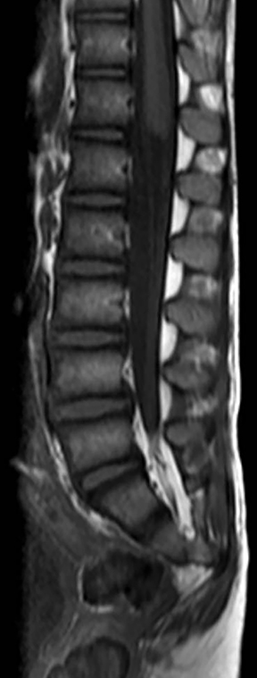
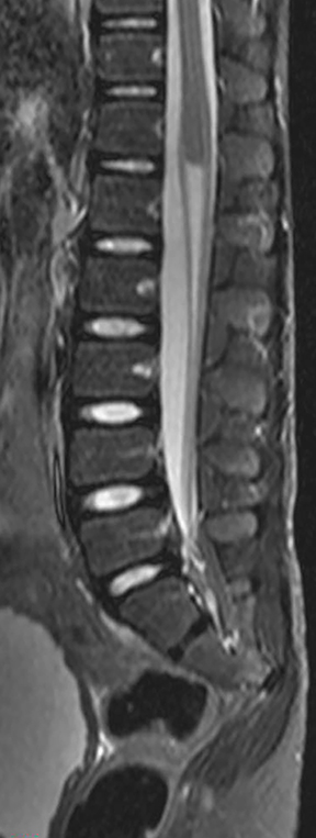
An otherwise asymptomatic 9-year-old girl was referred for pediatric neurology consultation for neurogenic bladder with urinary incontinence and chronic constipation. At observation she showed distal atrophy of the lower limbs and short gluteal cleft. She had had poor follow up over the years, and this was the first imaging exam she ever had, an MRI of the lumbar and sacral spine. Further investigation revealed that the child’s mother was diabetic.
IMAGING FINDINGS
MRI of the spine was performed with sagittal T1 and sagittal STIR. The images show partial sacrococcigeal agenesis with S2 being the last fully formed vertebra and with a hypoplastic S3. There is a complete absence of coccyx. There is also an abrupt wedge-shaped terminus of the cord, which lies opposite T12 and an abnormal course of the cauda equina with separation of the anterior and posterior nerve roots, forming the “double bundle shape.” The dural sac is abnormally highly placed, ending at L5.
DIAGNOSIS
Caudal regression syndrome type 1
DISCUSSION
Traditionally caudal agenesis/regression syndromes were classified according to the type of articulation between the ilia and the lowest vertebra present – Renshaw classification.1
More recently, MRI has allowed a better assessment of the spinal cord, allowing for a new classification based not on the bone defect but on the embryogenic error of the development of the spinal cord. Two types of caudal regression syndromes have been described:2
Type 1, in which not only the caudal tail bud is affected, but also part of the true notochord fails to develop (interference with both primary and secondary neurulation). There is aplasia of the caudal metameres with abrupt wedge-shaped terminus of the cord, which is high-lying.
Type 2, in which only the tail bud fails to develop and the true notochord remains unaffected (interference only with secondary neurulation). The vertebral anomaly is less severe and the conus is low and tethered.
An imaging evaluation is key to the diagnosis and, while evaluating the images, an assessment of the number of sacral vertebrae and their symmetry should be made. It is also important to evaluate the level and the shape of the cord terminus.
This syndrome is seen in up to 0.25/10,000 of normal pregnancies, but is reported to be more frequent in the offspring of diabetic mothers. It has a wide spectrum of clinical presentations depending on the severity of the caudal regression. The most common type, caudal regression syndrome type 1, is our case and is usually associated with motor impairment and deformities of the lower limbs.
The treatment depends, obviously, on the clinical presentation. In our case, the incontinence required a continence control system. In more severe cases, for example, an imperforated anus, a permanent colostomy is mandatory.
CONCLUSION
An imaging evaluation is key to the diagnosis of caudal regression syndrome and, while evaluating the images, an assessment of the number of sacral vertebrae and their symmetry should also be made. It is also important to evaluate the level and the shape of the cord terminus.
REFERENCES
- Kobayashi E, Setsu N. Osteosclerosis induced by denosumab. Lancet. 2014 Oct;pii: S0140-6736(14).
- Hakozaki M, Tajino T, Yamada H, Hasegawa O, Tasaki K, Watanabe K, Konno S. Radiological and pathological characteristics of giant cell tumor of bone treated with denosumab. Diagn Pathol. 2014;9:111.
- Brodowicz T, O’Byrne K, Manegold C. Bone matters in lung cancer. Ann Oncol. 2012;23(9): 2215-2222.
- Vallet S, Smith M, Raje N. Novel bone-targeted strategies in oncology. Clin Cancer Res. 2010;16(16):4084-4093.
- Kurata T, Nakagawa K. Efficacy and safety of denosumab for the treatment of bone metastases in patients with advanced cancer. Jpn J Clin Oncol.2012;42(8):663-669.
- Padhani AR, Gogbashian A. Bony metastases: Assessing response to therapy with whole-body diffusion MRI. Cancer Imaging. 2011;11 S129-145.
Citation
JB T, A B.Caudal regression syndrome. Appl Radiol. 2017; (1):47A-47B.
January 10, 2017