Automated ultrasound protocols enhance department efficiency
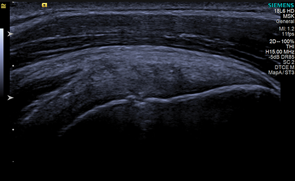
Images

Ultrasound is the most operator-dependent imaging modality. The skill of the operator in obtaining the images is a critical component in the radiologist’s ability to confidently provide a correct diagnosis. However, operator skill varies as a result of experience, education, and knowledge of the ultrasound system capabilities. This variability can lead to significant differences in the diagnostic images provided, even in same type of study or on the same patient. Notable differences are often gaps in anatomy between subsequent images, different views of a specific area of the anatomy, or simply a missed view/image altogether.
In 2007, an article in Radiology Management highlighted the benefits of protocol-driven ultrasound exams, citing a reduction in exam times and repetitive motion injuries, as well as greater consistency in acquired images which could enhance the quality of the study and diagnostic report.1
At Winthrop University Hospital (Mineola, NY), we provide general and vascular ultrasound imaging services through the radiology department and also at an external outpatient office. We pursued creating protocols for our ultrasound studies to address the issue of study variability by creating tabs that contained set protocols specific to an exam ordered on our existing ultrasound equipment. However, due to the age of the systems, we didn’t have the same flexibility provided by some newer systems that enable the programming of automated protocols by the type of ultrasound exam.
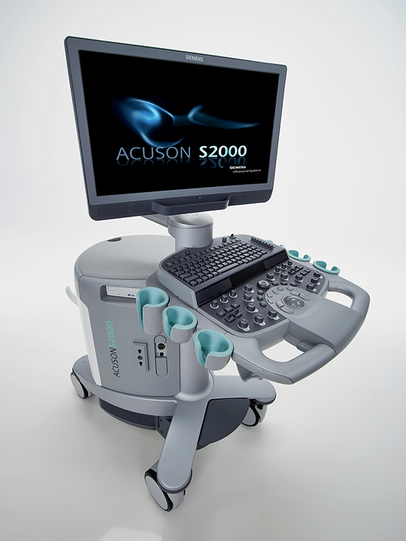
In February 2013, we replaced our 13-year old ultrasound systems with Siemens ACUSON systems: one S3000 system, four S2000 systems, and two S1000 systems, the later dedicated specifically for vascular use. These systems satisfied our two key requirements: the ability to fully implement automated protocols and generate digital dynamic clips, similar to “videos,” of the anatomy.
We worked closely with Pantea Karimi, BS, RDMS, RDCS, Clinical Sales Consultant, Siemens Healthcare, to initially set-up the eSieScan automated protocols that are available on the ACUSON S systems. As an ACR and IAC accredited facility, we reviewed practice guidelines to ensure our automated protocols not only met but exceeded these requirements. We also engaged our radiologists to determine the images they preferred.
The top priority was to develop a consistent workflow among our 20 sonographers for each and every ultrasound study. In general, every sonographer has a slightly different technique based on their education and clinical background; therefore, addressing this variability and creating uniformity in the ultrasound studies was important for our department.
Through the eSieScan protocols, we can specify the precise type and order of images the sonographer must capture for a particular study or anatomical area. The protocols include when to measure, utilize color and/or Doppler images and the angle of the Doppler. Once the protocol for each specific type of exam was finalized and tested, it was copied to a USB and then downloaded to each ultrasound system. The protocols can be created and revised on the S-Family ultrasound system or on a desktop PC.
Further, we began to investigate the use of digital dynamic clips feature, which captures live sweeps of the region of interest and helps eliminate “gaps” in the anatomy. A 2013 study published in the Journal of Ultrasound in Medicine reported that the use of digital dynamic clips increased the detection rate of fetal cardiac defects by up to 38%.2 Digital dynamic clips were included in the automated protocol implementation for each ultrasound exam.
Once our super user mastered the use of the automated protocols, other sonographers were trained. Implementing these protocol changes has been a seamless process with “buy-in” from all users and readers (radiologists). Our lead sonographer, Jamielee Prestano, BS, RDMS, RVT, continues to monitor the use of protocols and any changes from our regulatory bodies (ACR and IAC) so that we can constantly enhance them as needed.
More complete and consistent exams
As a result of implementing the eSieScan automated protocols, our sonographers are more efficient, our studies are more complete with no “missing” images, studies by body area/type are consistent across the machines/systems and sonographers, and our radiologists can anticipate the order and type of images in the study so they are also more efficient in their reading.
The consistency in our exams enables a common baseline in the provision of care for our patients receiving an ultrasound study. We’ve removed the variability in ultrasound imaging that will typically occur without a set parameter—this was not possible without the implementation of the automated protocols. That baseline is also important for follow-up studies, providing the radiologist with studies acquired in the same way, which enhances their ability to compare the anatomy or region of interest. Yet, there is flexibility for the sonographer to adapt to each individual case as needed by simply pausing the protocol.
Once the sonographers are trained on the use of automated protocols, scanning time per case decreases. With this decrease, we are able to accept the higher volume of cases being referred to our department and outpatient center—in 2014 our volume grew by approximately 8%.
As a sonographer sweeps through an area, the still image slices may not fully represent the anatomy or region of interest. With the digital dynamic clips as part of our protocol for all exams, we are capturing a more complete review of the anatomy in a consistent manner and our radiologists can view the cine clips along with the static images. While digital dynamic clips are not required by an accrediting organization at this time, we believe it is an important part of the study that helps eliminate gaps in the anatomy and provides the radiologists with a more comprehensive view of the patient’s injury or disease.
Joseph Mazzie, DO, a radiologist at Winthrop University Hospital, recently shared feedback on the changes we’ve implemented in ultrasound imaging. “Consistency is very important with regards to MSK work,” he says. “The automated protocols reduce the imaging time of patients in half and ensure that everything is covered—nothing is skipped. Additionally, the utilization of dual screen imaging with color and grayscale is extremely efficient.”
Dr. Mazzie reviews the clips for each study he interprets to ensure that he looked at all the images. “I find them very helpful for vascular and MSK studies, especially for studies done overnight when I’m not available to help ensure that all imaging views are acquired. We’ve found it also helps with reducing patient callbacks for additional imaging.”
Although the digital dynamic clips are now an important component of our ultrasound exams, the large file size has been challenging for our PACS. I would encourage other sites considering implementing this feature in ultrasound studies to investigate the requirements and capabilities of its PACS to ensure a more seamless storage, access and retrieval process.
Today at Winthrop University Hospital, we are providing consistent, high-quality ultrasound imaging across every exam as a result of implementing the eSieScan automated protocols and digital dynamic clips. Our sonographers and radiologists are more efficient, patient call backs due to missing images or views are significantly reduced, and our staff is providing a high level of clinical care utilizing some of the latest technologies in ultrasound imaging.
References
- Brandli L. Benefits of Protocol-Driven Ultrasound Exams. Radiology Management. 2007 Jan;29(4):56-59.
- Scott TE, Jones J, Rosenberg H. Increasing the detection rate of congenital heart disease during routine obstetric screening using cine loop sweeps. J Ultrasound Med. 2013 June;32(6):973-979.